


Pterotrachea scutata
Roger R. Seapy
Introduction
Pterotrachea scutata is immediately distinguished from the other two species in the genus by the lateral expansion of the anterior portion of the trunk, forming a large cuticular disk. The eyes are rectangular in dorsal view, very similar to those in P. coronata. The visceral nucleus is relatively short, tear-drop shaped, similar to that in P. hippocampus, and markedly shorter than in P. coronata. Unique to the species is a wreath of two or three rows of peribuccal teeth located inside the mouth opening. Morphology of the central rachidian tooth on the radula is similar to that in P. hippocampus. The larva is unknown, but is probably either larva 2 or 3 of Richter (1968, see Pterotrachea larvae). The geographical distribution is cosmopolitan in tropical to subtropical waters.
Characteristics
- Body morphology
- Anterior portion of the trunk greatly expanded, forming a cuticular disk, or bib (see title illustration)
- Proboscis narrow, cylindrical, and much shorter than in P. hippocampus or P. coronata; extending ventrally from beneath the anterior part of the cuticular disk, which overhangs the base of the proboscis
Click on an image to view larger version & data in a new window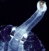
Figure. Dorsal view of the anterior part of the cuticular disk, with the proboscis extending ventrally from beneath the disk. ©
- Eyes rectangular in dorsal view, with the retinal base somewhat wider than the lens, as in P. coronata.
Click on an image to view larger version & data in a new window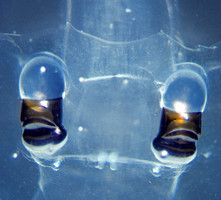
Click on an image to view larger version & data in a new windowFigure. Dorsal view of the eyes in Pterotrachea scutata. ©
- Visceral nucleus tear-drop shaped; similar to that in P. hippocampus, and markedly shorter than in P. coronata
Click on an image to view larger version & data in a new window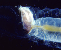
Figure. Lateral view of the tear-drop shaped visceral nucleus in Pterotrachea scutata. ©
- Swimming fin located on the middle of the trunk, but closer to the visceral nucleus than the eyes (see title illustration)
- Swimming fin sucker in males only; small and located anterior to the midpoint on the fin ventrum
Click on an image to view larger version & data in a new window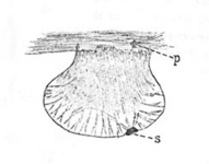
Figure. Swimming fin and sucker (=s) in Pterotrachea scutata. Also noted is the location of the pedal ganglia (=p). © 1949 J. J. Tesch
- Wreath of two or three irregular rows of tiny peribuccal teeth inside the entrance of the mouth; not present in other species of Pterotrachea. The peribuccal teeth were first characterized by Vayssière (1904) and subsequently remarked upon and illlustrated by Tesch (1949; see figure below)
Click on an image to view larger version & data in a new window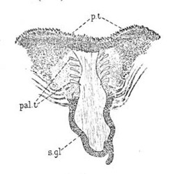
Figure. Drawing of the buccal cavity in Pterotrachea scutata, which was exposed by cutting along the ventral midline and removing the radula and its musculature. Immediately inside the mouth opening, the peribuccal teeth (p.t) form a ring, two or three rows deep. The much larger palatine teeth (pal.t) are located in the roof of the buccal cavity, along with the salivary glands (s.gl). © 1949 J. J. Tesch
- Larva unknown but represented, most probably, by either larva 2 or 3 of Richter (1968); see Pterotrachea larvae
- Radular morphology similar to that in the other species in the genus (see Pterotrachea radula), with a central tooth bearing a large, pointed medial cusp, flanked laterally by multiple short and pointed cusps.
Comments
Tesch (1949) reported that the lens in the eye of P. scutata was noticeably wider than that of P. coronata. Comparison of the eyes in a 61 mm P. scutata with those in a 78 mm P. coronata (Seapy, 1985, Fig. 3E,F) provide supportive evidence for Tesch's observation; despite the shorter body length of the P. scutata specimen, its lens width was greater (1.4 mm) than in P. coronata (1.1 mm).
The diel vertical distribution of Pterotrachea scutata is known only from results of a study by Pafort-van Iersel (1983) that was based on samples collected from the mid North Atlantic Ocean using paired 8-m2 and 1-m2 opening-closing, rectangular midwater trawls. Among the three species of Pterotrachea, the number of P. scutata (74) collected was comparable to P. hippocampus (72), but only one-half as many as P. coronata (146). The numbers of P. scutata collected by the paired nets were markedly different; all but 5 were recorded from the larger net. Based on the difference in the areas of the mouth openings of the two nets, one would predict about an 8-fold difference in the numbers captured by the two nets; instead there was a 15-fold difference. Alternatively, the difference could be explained by an ability of P. scutata to largely avoid capture by the smaller net. Related to this result was that nearly three times as many individuals were collected at night (47) than during the day (17). Since heteropods have image-forming eyes and are visual predators, a reasonable explanation for this diel difference in numbers captured is the ability of the animals to visually detect and avoid net capture during the day. Vertical distribution data were obtained from six depth intervals; 50-100 m, at 100 m depth intervals between 100 and 500 m, and 500-1,000 m. From the daytime tows, only three individuals were captured at 100-200 m, none from 200-300 m, and fifteen below 300 m. In contrast, 39 specimens were collected between 100 and 300 m at night and only eight below 300 m. These results support an hypothesis of nocturnal vertical migration from mesopelagic depths during day into the epipelagic zone at night. Also, the absence of individuals from the six tows taken at 50-100 m during both day and night periods suggests that the species may not dwell in the upper 100 m.
References
Lalli, C. M. and R. W. Gilmer. 1989. Pelagic snails. The biology of holoplanktonic gastropod mollusks. Stanford University Press, Stanford. 259 pp.
Pafort-van Iersel, T. 1983. Distribution and variation of Carinariidae and Pterotracheidae (Heteropoda, Gastropoda) of the Amsterdam Mid North Atlantic Plankton Expedition 1980. Beaufortia 33: 73-96.
Richter, G. 1968. Heteropoden und Heteropodenlarven im Oberflächenplankton des Golfs von Neapel. Pubblicazioni della Stazione Zoologica di Napoli 36: 346-400.
Richter, G. and R. R. Seapy. 1999. Heteropoda, pp. 621-647. In: D. Boltovskoy (ed.), South Atlantic Zooplankton. Backhuys Publishers, Leiden.
Seapy, R.R. 1985. The pelagic genus Pterotrachea (Gastropoda: Heteropoda) from Hawaiian waters: a taxonomic review. Malacologia 26(1-2): 125-135.
Tesch, J. J. 1949. Heteropoda. Dana Report 34, 53 pp, 5 plates.
Vayssière, A. 1904. Mollusques hétéropodes provenant des campagnes des yachts Hirondelle et Princesse Alice. Résultats des campagnes scientifiques accomplies su son yacht par Albert 1er prince souverain de Monaco. 26: 3-65.
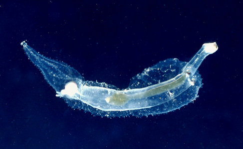
Title Illustrations

| Scientific Name | Pterotrachea scutata |
|---|---|
| Location | Hawaiian waters |
| Sex | Female |
| Life Cycle Stage | adult |
| View | ventral |
| Image Use |
 This media file is licensed under the Creative Commons Attribution-NonCommercial License - Version 3.0. This media file is licensed under the Creative Commons Attribution-NonCommercial License - Version 3.0.
|
| Copyright |
©

|
About This Page
California State University, Fullerton, California, USA
Correspondence regarding this page should be directed to Roger R. Seapy at
Page copyright © 2008
 Page: Tree of Life
Pterotrachea scutata .
Authored by
Roger R. Seapy.
The TEXT of this page is licensed under the
Creative Commons Attribution License - Version 3.0. Note that images and other media
featured on this page are each governed by their own license, and they may or may not be available
for reuse. Click on an image or a media link to access the media data window, which provides the
relevant licensing information. For the general terms and conditions of ToL material reuse and
redistribution, please see the Tree of Life Copyright
Policies.
Page: Tree of Life
Pterotrachea scutata .
Authored by
Roger R. Seapy.
The TEXT of this page is licensed under the
Creative Commons Attribution License - Version 3.0. Note that images and other media
featured on this page are each governed by their own license, and they may or may not be available
for reuse. Click on an image or a media link to access the media data window, which provides the
relevant licensing information. For the general terms and conditions of ToL material reuse and
redistribution, please see the Tree of Life Copyright
Policies.
- First online 17 September 2008
- Content changed 17 September 2008
Citing this page:
Seapy, Roger R. 2008. Pterotrachea scutata . Version 17 September 2008 (under construction). http://tolweb.org/Pterotrachea_scutata/28740/2008.09.17 in The Tree of Life Web Project, http://tolweb.org/


