


Pterotracheidae
Roger R. Seapy
- Firoloida desmarestia Lesueur 1817
- Pterotrachea Niebuhr (in Forskål) 1775
Introduction
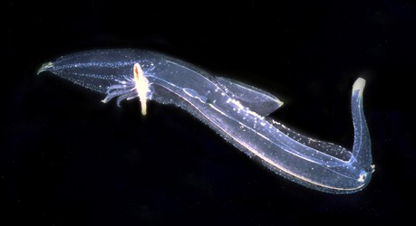
All pterotracheids lack a shell as adults, although they possess one as larvae (the shell is shed following metamorphosis). Their bodies are elongate and basically cylindrical, consisting of a proboscis, trunk and tail. The maximal recorded body length is 38.5 cm (Pterotrachea coronata). The head lacks tentacles anterior to the eyes, except in male Firoloida demarestia. The viscera are compacted into a long, cylindrical visceral nucleus. The swimming fin is large, located about midway between the anterior and posterior ends of the trunk, and bears a sucker only in males. Pterotracheids are mostly epipelagic (dwelling in the upper several hundred meters of the water column), although the vertical ranges of two species of Pterotrachea extend into the mesopelagic zone. All pterotracheids have cosmopolitan distributions in tropical to subtropical waters.
The Pterotracheidae are widely regarded as the most highly derived of the heteropod families. Features supporting this contention include: (1) enlargement, elongation and narrowing (to a basically cylindrical shape) of the body in the anterior-posterior axis, resulting in a streamlined body with enhanced swimming abilities, (2) shedding of the larval shell following metamorphosis, with the result that buoyancy problems are reduced since a calcareous shell (present in the adults of the other two families) is lacking, (3) compaction of the viscera into a pyriform visceral nucleus, which is largely enveloped by the gelatinous body at the posterior end of the trunk.
Brief Diagnosis
Heteropod molluscs with:
- Body elongated and basically cylindrical, divided into proboscis, trunk and tail
- Viscera compacted into a pyriform visceral nucleus
- Shell lacking in adults; larval shell shed after metamorphosis
- All species with cosmopolitan distributions in tropical to subtropical waters
Characteristics
- Shell present in larvae only; cast off following metamorphosis
- Body morphology
- Elongate, basically cylindrical; streamlined for rapid swimming
- Proboscis, trunk and tail regions well developed Click on an image to view larger version & data in a new window
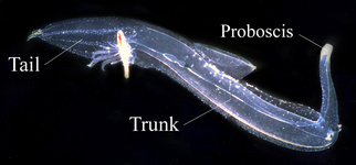
Figure. Body regions in Pterotrachea coronata. © R. R. Seapy
- Viscera compacted into a long and narrow visceral nucleus
- Esophagus extends from the buccal mass, at the end of the proboscis, to the visceral nucleus
- Swimming fin
- Located about midway between head region and visceral nucleus
- Fin sucker small; present only in malesClick on an image to view larger version & data in a new windowClick on an image to view larger version & data in a new windowClick on an image to view larger version & data in a new window
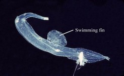

Figure. Pterotrachea coronata. Left: swimming fin in female, viewed from right side of body. Right: swimming fin and sucker in male, viewed from right side. © R. R. Seapy
- Located about midway between head region and visceral nucleus
- Head
- In dorsal view, shape of eyes either rectangular (with a narrow retinal base) or triangular (with a broad retinal base that curves upward toward the lens)
- Tentacles absent, except in Firoloida demarestia males
Click on an image to view larger version & data in a new windowClick on an image to view larger version & data in a new window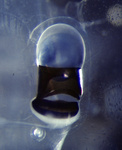
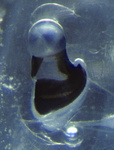
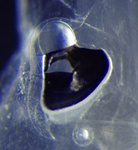
Figure. Dorsal views of eyes in Pterotrachea. Left: rectangular eye of P. coronata. Center: narrowly triangular eye in juvenile P. hippocampus. Right: broadly triangular eye in adult P. hippocampus. © R. R. Seapy
- In dorsal view, shape of eyes either rectangular (with a narrow retinal base) or triangular (with a broad retinal base that curves upward toward the lens)
- Radula
- 24-30 tooth rows
- Central rachidian tooth (lower center in photo below) polycuspid with a prominent middle cusp that is flanked on either side by numerous short cusps
Click on an image to view larger version & data in a new windowClick on an image to view larger version & data in a new window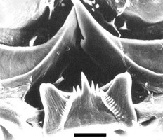
Figure. High magnification view of central rachidian tooth (lower, middle) and elongate, pointed lateral teeth (upper left and right in photo) from radula of Pterotrachea hippocampus. Scale bar = 20 µm. © G. Richter
Comments
Two genera are included in the Pterotracheidae, one of which, Firoloida, is monotypic. The genera can be distinguished by the following characters:
Genus Tail Tentacles anterior to the eyes
Posterior egg string
Pterotrachea well developed
absent in both sexes
absent Firoloida very short, ventral present in both sexes
present
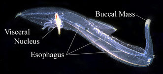
Figure. Location of buccal mass, esophagus, and visceral nucleus in Pterotrachea coronata. © R. R. Seapy
References
Lalli, C. M. and R. W. Gilmer. 1989. Pelagic snails. The biology of holoplanktonic gastropod mollusks. Stanford University Press, Stanford. 259 pp.
Richter, G. and R. R. Seapy. 1999. Heteropoda, pp. 621-647. In: D. Boltovskoy (ed.), South Atlantic Zooplankton. Backhuys Publishers, Leiden.
Spoel, S. van der, L. Newman and K. W. Estep. 1997. Pelagic molluscs of the world. World Biodiversity Database, CD-ROM Series. Expert Center for Taxonomic Identification (ETI), University of Amsterdam. UNESCO Publishing, Paris.
Tesch, J. J. 1949. Heteropoda. Dana Report 34, 53 pp., 5 plates.
Title Illustrations

| Scientific Name | Pterotrachea coronata |
|---|---|
| Location | Hawaiian Islands |
| Specimen Condition | Live Specimen |
| Sex | Female |
| Life Cycle Stage | adult |
| View | left side |
| Image Use |
 This media file is licensed under the Creative Commons Attribution-NonCommercial License - Version 3.0. This media file is licensed under the Creative Commons Attribution-NonCommercial License - Version 3.0.
|
| Copyright |
©

|
About This Page
California State University, Fullerton, California, USA
Correspondence regarding this page should be directed to Roger R. Seapy at
Page copyright © 2006
 Page: Tree of Life
Pterotracheidae .
Authored by
Roger R. Seapy.
The TEXT of this page is licensed under the
Creative Commons Attribution License - Version 3.0. Note that images and other media
featured on this page are each governed by their own license, and they may or may not be available
for reuse. Click on an image or a media link to access the media data window, which provides the
relevant licensing information. For the general terms and conditions of ToL material reuse and
redistribution, please see the Tree of Life Copyright
Policies.
Page: Tree of Life
Pterotracheidae .
Authored by
Roger R. Seapy.
The TEXT of this page is licensed under the
Creative Commons Attribution License - Version 3.0. Note that images and other media
featured on this page are each governed by their own license, and they may or may not be available
for reuse. Click on an image or a media link to access the media data window, which provides the
relevant licensing information. For the general terms and conditions of ToL material reuse and
redistribution, please see the Tree of Life Copyright
Policies.
- First online 30 May 2006
- Content changed 14 August 2008
Citing this page:
Seapy, Roger R. 2008. Pterotracheidae . Version 14 August 2008 (under construction). http://tolweb.org/Pterotracheidae/28734/2008.08.14 in The Tree of Life Web Project, http://tolweb.org/


