- Mantle
- Mantle opening narrow.
 Click on an image to view larger version & data in a new window
Figure. Enlargement of one of the title photographs of L. shuishi showing the funnel and mantle opening. Photograph from O'Shea and Lu, 2002.
- Optic lobe
- Optic lobe spherical.
- Single optic nerve bundle arises from optic lobe.
Click on an image to view larger version & data in a new window
Figure. Dissection of the head of L. shuishi showing optic lobe, nerve bundle and white body of holotype. Photograph from O'Shea and Lu, 2002.
- Beaks
- Lateral walls of lower beaks with a weak fold on either side that runs from the jaw angle to the lower corner of lateral wall.
Click on an image to view larger version & data in a new window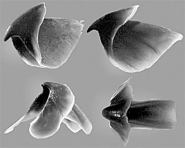
Figure. Oblique and lateral views of the upper beak (top) and the same views of the lower beak (bottom) of L. shuishi, holotype. Photographs from O'Shea and Lu, 2002.
- Radula
- Radula with 8 rows of long narrow, homodont teeth.
Click on an image to view larger version & data in a new window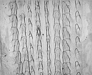
Figure. Radula of L. shuishi, holotype. Note the asymmetry in the photograph. A diamond shape is placed on the rhachidian tooth series. The extra tooth series (coleoid radulas have a maximum of seven series), apparently, represents an enlarged marginal plate along the right side. Photomicrograph from O'Shea and Lu, 2002
- Pigmentation
- Dorsal and ventral surfaces of mantle, head and arms white to translucent.
- Distal margin of fin pinkish red (see dorsal and ventral views of the web in photographs below) .
- Oral face of arms beside each sucker row pinkish red as is central, oral region of the web.
- Cirri darker red.
- Eyes scarlet, opening to eye (eyelid) lightly pigmented (see arrow in the photograph).
- Tip of funnel heavily pigmented (see photograph at the top of this page).
Click on an image to view larger version & data in a new window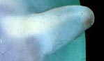
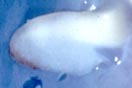
Figure. Dorsal (left) and ventral (right) views of the fins of L. shuishi, holotype. Photographs from O'Shea and Lu, 2002.
Click on an image to view larger version & data in a new window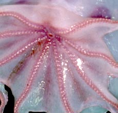
Figure. Oral view of arms and web of L. shuishi, holotype. Photograph from O'Shea and Lu, 2002.
Click on an image to view larger version & data in a new window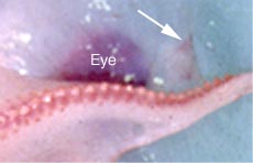
Figure. Enlargement of portion of the head of L. shuishi showing eye and opening of canal that leads to the eye, holotype. Photograph from O'Shea and Lu, 2002.
- Measurements and counts
* DamagedSex Female Mantle length 87.5 mm Mantle width 130 Mantle aperature 30 Fin length 95 Fin width 48 Eye lid opening 8 Arm I, length (right/left) 250*/238 Arm II, length 280/259 Arm III, length 278/244 Arm IV, length 223/242 Web depth, sector A 67.5 Web depth, sector B 53/60 Web depth, sector C 51/68 Web depth, sector D 39/50 Web depth, sector E 30 Arm I, sucker count 47*/35* Arm II, sucker count 57*/44* Arm III, sucker count 46*/43* Arm IV, sucker count 36*/40* Max. cirrus length 1.0-1.5 Max. sucker diameter 3.7-4.0 Gill lamellae - These measurements and counts are from O'Shea and Lu, 2002.
Luteuthis shuishi Description Continued
Steve O'Shea, Chung Cheng Lu, Richard E. Young, and Michael VecchioneReferences
O'Shea, S. and C. C. Lu. 2002. A New Species of Luteuthis (Mollusca: Cephalopoda: Cirroctopoda) from the South China Sea. Zoological Studies, 41: 119-126.
About This Page
Steve O'Shea

Oceanic Sciences Research Institute, Auckland University of Technology, Auckland, New Zealand
National Chung Hsing University Taiwan
University of Hawaii, Honolulu, HI, USA
National Museum of Natural History, Washington, D. C. , USA
Page copyright © 2003 , , , and C. C. Lu
All Rights Reserved.


