


Cranchia
Cranchia scabra
Richard E. Young and Katharina M. Mangold (1922-2003)
Introduction

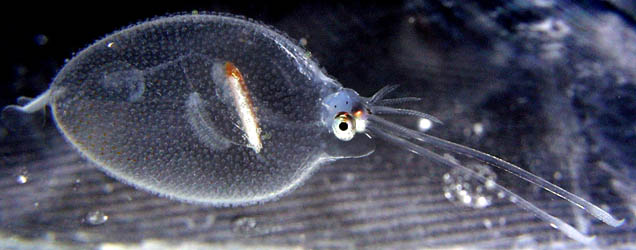
Figure. C. scabra at 532 m. © MBARI 2013
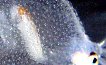
Figure. Lateral view of part of the mantle and head of a 30 mm ML C. scabra showing tubercles. Photograph by R. Young.
C. scabra, the only species in the genus, is small (150 mm ML) and one of the most distinctive cranchiids.
The mantle is covered by large, multi-pointed cartilagenous tubercles (see Roper and Lu 1990, for a description of the tubercle structure). When disturbed, the squid often pulls its head and arms into the mantle cavity and folds its fins tightly against the mantle to form a turgid ball. The tubercules, presumably, provide some type of protection but it is unclear what predators are affected and how. In addition, the squid may ink into the mantle cavity, making the ball opaque. This was thought to be an aberrant behavior due to stress and confinement of shipboard aquaria until the same inking behavior was seen in cranchiids from submersibles (Hunt, 1996). The function of this behavior is unknown.
Brief diagnosis:
A cranchiin ...
- with mantle covered with cartilagenous tubercules.
Characteristics
- Tentacles
- Suckers in a transverse row on club manus of equal size.
- Diagonally set pairs of suckers and pad on distal 2/3 of tentacular stalk.
- Head
- Eyes sessile in paralarvae.
- Beaks: Descriptions can be found here: Lower beak; upper beak.
- Funnel
- Funnel valve present, large.
- Mantle
- Mantle covered with cartilagenous tubercles bearing 3-5 sharp cusps.
- Fins
- Each fin nearly oval in shape with free posterior lobes. Click on an image to view larger version & data in a new window

Figure. Ventral view of the fins of C. scabra. 120 mm ML. Drawing from N. Voss, 1980, p. 377.
- Each fin nearly oval in shape with free posterior lobes.
- Photophores
- Fourteen oval photophores on each eye (ventral-proximal series of 8 photophores, ventral-distal series of 4 photophores near lens and dorsal series of two photophores near lens). Click on an image to view larger version & data in a new window
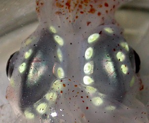
Figure. Ventral view of most ocular photophores of C. scabra. The dorsal series of photophores is not visible. Photograph by M. Vecchione.
- Photophores on tips of all arms in mature or nearly mature females.
Click on an image to view larger version & data in a new window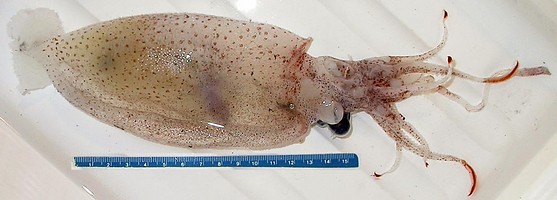
Figure. Ventral view of a maturing (?) but damaged female C. scabra, Gulf of Mexico, 150 mm ML, showing the red-pigmented photophores at the tips of the arms. Photograph by M. Vecchione.
Comments
Characteristics are from Voss (1980). More details of the description of C. scabra can be found here.
Behavior
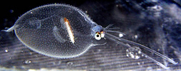
The small C. scabra below, photographed in a shipboard aquarium, has retracted its head with arms and tentacles into the mantle cavity. The mantle has taken the shape of a sphere and the chromatophores have expanded. This response to disturbance perhaps makes their consumption by small-mouthed predators more difficult.
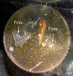
Figure. Posterolateral view of C. scabra, 30 mm ML. Photograph by R. Young.
The photographs below, also taken in an aquarium, show two different color phases of the same squid. A typical transparent phase on the right and a peculiar anteriorly pigmented phase on the left. The half-pigmented phase was seen several times (D. Fenolio, pers. communication).
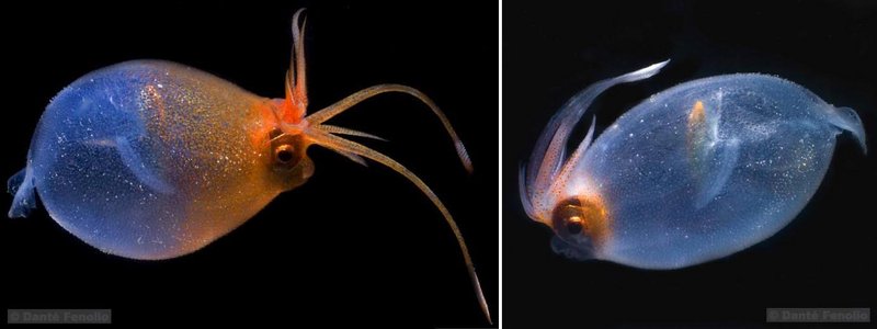
Figure. Two side views of the same C. scabra, Sea of Japan, ca. 10 cm ML. Photographs by Danté Fenolio.
Life History
Small paralarvae lack tubercles and are similar in appearance to paralarvae of Liocranchia. However they can easily be separated from paralarvae of Liocranchia by the numerous, scattered chromatophores that cover much or all of the mantle and by the short, thick tentacles that appear to be rugose or, possibly, glandular. Note the sessile eyes. By 8 mm ML they have numerous tubercules.
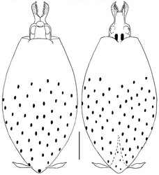
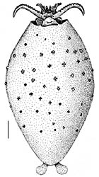
Figure. Paralarval C. scabra. Left - Ventral and dorsal views, 4.7 mm ML, Hawaiian waters. Chromatophores were faint and pattern may not be complete; at slightly larger sizes the chromatophores cover the entire mantle. Drawings by R. Young. Right - Ventral view, 8 mm ML, showing tubercules, not chromatophores. Drawing from Voss, 1980, p. 377, printed with the permission of the Bulletin of Marine Science. Scale bar is 1 mm.
Distribution
This species occurs throughout tropical and subtropical waters of the world's oceans (Nesis, 1982).
References
Hunt, J. 1996. The behavior and ecology of midwater cephalopods from Monterey Bay: Submersible and laboratory observations. Doctoral Diss., Univ. Calif. Los Angeles.
Roper, C. F. E. and C. C. Lu 1990. Comparative morphology and function of dermal structures in oceanic squids (Cephalopoda). Smithson. Contr. Zool., No. 493: 1-40.
Young, R. E. 1972. The systematics and areal distribution of pelagic cephalopods from the seas off Southern California. Smithson. Contr. Zool., 97: 1-159.
Title Illustrations
| Scientific Name | Cranchia scabra |
|---|---|
| Comments | photographed in a shipboard aquarium off Hawaii. |
| Size | 30 mm ML |
| Image Use |
 This media file is licensed under the Creative Commons Attribution-NonCommercial License - Version 3.0. This media file is licensed under the Creative Commons Attribution-NonCommercial License - Version 3.0.
|
| Copyright |
© 1998

|
About This Page
University of Hawaii, Honolulu, HI, USA
Katharina M. Mangold (1922-2003)
Laboratoire Arago, Banyuls-Sur-Mer, France
Page copyright © 2019 and Katharina M. Mangold (1922-2003)
 Page: Tree of Life
Cranchia . Cranchia scabra .
Authored by
Richard E. Young and Katharina M. Mangold (1922-2003).
The TEXT of this page is licensed under the
Creative Commons Attribution-NonCommercial License - Version 3.0. Note that images and other media
featured on this page are each governed by their own license, and they may or may not be available
for reuse. Click on an image or a media link to access the media data window, which provides the
relevant licensing information. For the general terms and conditions of ToL material reuse and
redistribution, please see the Tree of Life Copyright
Policies.
Page: Tree of Life
Cranchia . Cranchia scabra .
Authored by
Richard E. Young and Katharina M. Mangold (1922-2003).
The TEXT of this page is licensed under the
Creative Commons Attribution-NonCommercial License - Version 3.0. Note that images and other media
featured on this page are each governed by their own license, and they may or may not be available
for reuse. Click on an image or a media link to access the media data window, which provides the
relevant licensing information. For the general terms and conditions of ToL material reuse and
redistribution, please see the Tree of Life Copyright
Policies.
- Content changed 26 March 2019
Citing this page:
Young, Richard E. and Katharina M. Mangold (1922-2003). 2019. Cranchia . Cranchia scabra . Version 26 March 2019. http://tolweb.org/Cranchia_scabra/19542/2019.03.26 in The Tree of Life Web Project, http://tolweb.org/


