


Carinaria cristata
Roger R. Seapy

Introduction
Carinaria cristata attains the largest body size (≥ 680 mm body length) of all heteropods. The proboscis is short and the trunk inflated. The tail is larger (in proportion to the trunk and proboscis) than in any other species of Carinaria, with a tall dorsal crest that slopes posteriorly and leads to a whip-like posterior portion. In large adults the dorsal surface of the tail develops a pattern of brown pigmentation and the posterior part of the tail is brown. The shell is triangular in side view, with a height to basal length ratio of 0.8-1.0. With increasing size, the apical portion of the shell slopes posterioly. The keel is very low, increasing gradually in height towards the shell aperture.
Brief Diagnosis
A species of Carinaria with the following characteristics:
- Reaches the largest body size among all the species of heteropods (≥680 mm)
- Proboscis short and trunk inflated
- Tail very large and elongate, with a tall dorsal crest
- In large animals, dorsal surface of tail with distinct pattern of brown pigmentation
- Shape of shell triangular in side profile, with a very low keel
Characteristics
- Body morphology
- Proboscis short; trunk inflated with a thick cutis
- Viewed dorsally, eye shape triangular
- Tentacles present anterior to eyes; right one smaller than the left in older individuals, but in younger animals the rignt one is somewhat longer than the left (Tesch, 1949)
- Visceral nucleus stalk short, with the result that the visceral mass and shell are positioned lower than in other species of Carinaria
- Tail very large and elongate; length greater than that of the combined proboscis and trunk (to 175%; see figure below)
- Tail crest tall, tapering posteriorly to a whip-like posterior region that is triangular in cross-section
- In older individuals, dorsal surface of tail crest brown with short, ventrally-directed brown bands, and posterior section of tail brown (contrast the tails of the animals in the title illustration and in the figure below)
Click on an image to view larger version & data in a new window
Figure. Largest specimen (body length ≥ 680 mm) of Carinaria cristata recorded to date. Proboscis and trunk of specimen appear to be contracted; thus, the actual body length is undoubtedly greater than 680 mm. Specimen missing its visceral nucleus and shell at the time of collection by hand from the ocean surface outside Palmyra Atoll. © 2007 John Svenson
- Proboscis short; trunk inflated with a thick cutis
- Shell morphology
- In side view shell triangular, with apical part of the shell sloping posteriorly. Height to basal length ratio of shell = 0.8-1.0 (Okutani, 1961)
- Keel very low and increases gradually in height with proximity to shell aperture (see middle photograph below)Click on an image to view larger version & data in a new windowClick on an image to view larger version & data in a new window

Figure. Three shells of Carinaria cristata of decreasing size. Apical portion of shell on left missing. Photographs from Tesch (1906, Plate 9, Figs. 38-40). Scale bars based on shell sizes given in figure legends. © 1906 J. J. Tesch
Click on an image to view larger version & data in a new window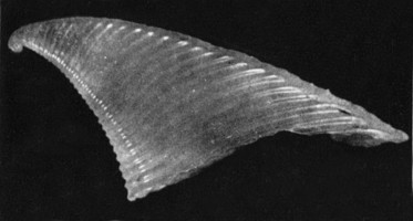
Figure. Shell of Carinaria cristata, viewed from right side. Shell height = 22.6 mm. © 1961 Takashi Okutani
- In side view shell triangular, with apical part of the shell sloping posteriorly. Height to basal length ratio of shell = 0.8-1.0 (Okutani, 1961)
- Radular morphology with a central (rachidian) tooth bearing three pointed cusps; typical of carinariids
Click on an image to view larger version & data in a new window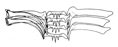
Figure. Drawing of a section of the radula in Carinaria cristata. To distinguish between the shapes of the two marginal and the single lateral teeth that are present on both sides of the central tooth in each tooth row, marginal teeth are illustrated on the left and lateral teeth on the right of the central tooth row. © 1976 S. van der Spoel
Click on an image to view larger version & data in a new window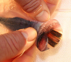
Figure. Anterior end of proboscis with the mouth held open to show the radula. The lighter- colored central teeth are flanked, on either side, by the darker-colored, overlapping marginal and lateral teeth. © 2007 John Svenson
References
Okutani, T. 1961. Notes on the genus Carinaria (Heteropoda) from Japanese and adjacent waters. Publications of the Seto Marine Biological Laboratory 9: 333-352.
Spoel, S. van der. 1976. Pseudothecosomata, Gymnosomata and Heteropoda (Gastropoda). Bohn, Scheltema and Holkema, Utrecht. 484 pp.
Tesch, J. J. 1906. Die Heteropoden der Siboga-Expedition. Monograph 51, 112 pp, 14 plates. E. J. Brill, Leiden.
Tesch, J. J. 1949. Heteropoda. Dana Report 34, 53 pp., 5 plates.
Title Illustrations

| Scientific Name | Carinaria cristata |
|---|---|
| Comments | In the upper animal, the visceral nucleus and shell are missing; presumably eaten by a predator. The smaller animal below lacks the brown coloration on the tail and has an intact shell that is curiously positioned low on the trunk |
| Reference | Tesch, J. J. 1906. Die Heteropoden der Siboga Expedition. Monographie 51, 112 pp. and 14 plates. |
| Specimen Condition | Dead Specimen |
| Sex | female (above) and male? (below) |
| Life Cycle Stage | adult |
| View | right side |
| Size | body length = ca. 235 mm (top) and 175 mm (bottom) |
| Copyright | © 1906 J. J. Tesch |
About This Page

California State University, Fullerton, California, USA
Correspondence regarding this page should be directed to Roger R. Seapy at
Page copyright © 2007
 Page: Tree of Life
Carinaria cristata .
Authored by
Roger R. Seapy.
The TEXT of this page is licensed under the
Creative Commons Attribution License - Version 3.0. Note that images and other media
featured on this page are each governed by their own license, and they may or may not be available
for reuse. Click on an image or a media link to access the media data window, which provides the
relevant licensing information. For the general terms and conditions of ToL material reuse and
redistribution, please see the Tree of Life Copyright
Policies.
Page: Tree of Life
Carinaria cristata .
Authored by
Roger R. Seapy.
The TEXT of this page is licensed under the
Creative Commons Attribution License - Version 3.0. Note that images and other media
featured on this page are each governed by their own license, and they may or may not be available
for reuse. Click on an image or a media link to access the media data window, which provides the
relevant licensing information. For the general terms and conditions of ToL material reuse and
redistribution, please see the Tree of Life Copyright
Policies.
- First online 12 July 2008
- Content changed 12 July 2008
Citing this page:
Seapy, Roger R. 2008. Carinaria cristata . Version 12 July 2008 (under construction). http://tolweb.org/Carinaria_cristata/28748/2008.07.12 in The Tree of Life Web Project, http://tolweb.org/


