


Carinaria cithara
Roger R. Seapy
Introduction
Two morphological features, eye and shell shape, distinguish Carinara cithara from the other species in the genus. In side profile the shell is tall and narrow, and in adults it has the greatest height to basal length ratio (2.0 or higher) of all Carinaria. The shape of the eye, when viewed dorsally, is cylindrical due to the narrow retinal base that is only slightly wider than the lens. In all other species in the genus the eye shape is triangular because the retinal base is much wider than the lens. The protoconch of the shell is prominently located at the top of the shell; second whorl of protoconch with a distinctive pattern of spiral sculpture. The tail is large, with a low dorsal crest.
Brief Diagnosis
A species of Carinaria with:
- Body length to 50 mm
- Shell shape, viewed from the side, is tall and narrow (height to basal length ratio ≥ 2.0)
- Protoconch positioned dorsally on shell apex, with spiral sculpture on second whorl
- Shell keel moderately low and uniform in height
- Tail large with a low doral crest
Characteristics
- Body morphology
- Body shape cylindrical; transition from proboscis to trunk to anterior part of tail not strongly demarcated
- Eyes cylindrical in dorsal view, with retinal base only slightly wider than the lens
- Tail relatively large, with a low dorsal crest
Click on an image to view larger version & data in a new window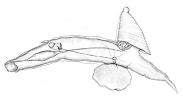
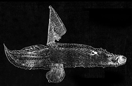
Figure. Drawing of young Carinaria cithara, from the left side. From the low height to basal length ratio of the shell, this is presumably a young individual. © J. A. McGowan
- Shell morphology
- Shell laterally compressed and tall; height to basal length ratio ≥ 2.0 (in large animals, see right side of first figure below). In small animals, the ratio may approach 1.0 (see second figure below)
- Apical region dorsal on shell; protoconch (larval shell) about 1.0 mm diameter; second whorl with spiral ridges (see left side of first figure below)
Click on an image to view larger version & data in a new window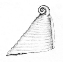
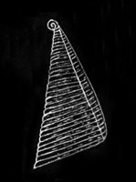
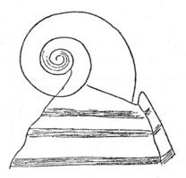
Figure. Shell of Carinaria cithara, viewed from right side. Left: shell from young individual © J. A. McGowan. Middle: adult shell. Right: apical region of middle shell. © J. J. Tesch
- Radula
- Central rachidian teeth broad, with three median cusps and two lateral cusps (below left). Median cusps long and pointed, with basal parts elevated and thick; presumably adding strength to each cusp (below right). Lateral cusps short and hooked inwards (below left)Click on an image to view larger version & data in a new window
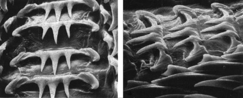
Figure. Central rachidian teeth on Carinaria cithara radula. Left: dorsal view. Right: view at tilted angle. © 1973 Catherine Thiriot
- Central rachidian teeth broad, with three median cusps and two lateral cusps (below left). Median cusps long and pointed, with basal parts elevated and thick; presumably adding strength to each cusp (below right). Lateral cusps short and hooked inwards (below left)
References
Okutani, T. 1961. Notes on the genus Carinaria (Heteropoda) from Japanese and adjacent waters. Publications of the Seto Marine Biological Laboratory 9: 333-352.
Spoel, S. van der. 1976. Pseudothecosomata, Gymnosomata and Heteropoda (Gastropoda). Bohn, Scheltema and Holkema, Utrecht. 484 pp.
Tesch, J. J. 1906. Die Heteropoden der Siboga-Expedition. Monograph 51, 112 pp, 14 plates. E. J. Brill, Leiden.
Tesch, J. J. 1949. Heteropoda. Dana Report 34, 53 pp., 5 plates.
Thiriot-Quiévreux, C. 1973. Observations de la radula des Hétéropodes (Mollusca Prosobranchia) au microscope électronique à balayage et interprétation fonctionnelle. Comptes rendus de l'Académie des Sciences, Serie D 276: 761-764.
Title Illustrations

| Scientific Name | Carinaria cithara |
|---|---|
| Sex | Female |
| Life Cycle Stage | adult |
| View | right side |
| Size | body length = 48 mm |
| Copyright | © 1949 J. J. Tesch |
About This Page

California State University, Fullerton, California, USA
Correspondence regarding this page should be directed to Roger R. Seapy at
Page copyright © 2007
 Page: Tree of Life
Carinaria cithara .
Authored by
Roger R. Seapy.
The TEXT of this page is licensed under the
Creative Commons Attribution License - Version 3.0. Note that images and other media
featured on this page are each governed by their own license, and they may or may not be available
for reuse. Click on an image or a media link to access the media data window, which provides the
relevant licensing information. For the general terms and conditions of ToL material reuse and
redistribution, please see the Tree of Life Copyright
Policies.
Page: Tree of Life
Carinaria cithara .
Authored by
Roger R. Seapy.
The TEXT of this page is licensed under the
Creative Commons Attribution License - Version 3.0. Note that images and other media
featured on this page are each governed by their own license, and they may or may not be available
for reuse. Click on an image or a media link to access the media data window, which provides the
relevant licensing information. For the general terms and conditions of ToL material reuse and
redistribution, please see the Tree of Life Copyright
Policies.
- First online 13 July 2008
- Content changed 22 July 2008
Citing this page:
Seapy, Roger R. 2008. Carinaria cithara . Version 22 July 2008 (under construction). http://tolweb.org/Carinaria_cithara/28747/2008.07.22 in The Tree of Life Web Project, http://tolweb.org/


