On this page we include beaks that don't fall into any of the three major beak types. These include: Ancistroteuthis (Onychoteuthidae), Ancistrocheiridae, Cycloteuthidae, the lepidoteuthid families (Lepidoteuthidae, Octopoteuthidae and Pholidoteuthidae) and Sepiidae.
ORDER OEGOPSIDA:
ONYCHOTEUTHIDAE: Ancistroteuthis lichtensteini
UPPER BEAK CHARACTERISTICS
- Step: Present. Between Wedge and Jaw Edge.
- Shoulder Blade: Present on outer part of Shoulder. Appears, however, to be covered, in some specimens, by a thin, partially translucent white/yellow layer thickest dorsally (Cover not seen at 107 mm ML); somewhat similar to that of other onychoteuthids.
- Shoulder Blade: Consists of one major component with a Wedge located anterior to it.
- Hyaline Matrix: Forms middle layer of Shoulder.
- Palatoshoulder Ridge: Present but greatly reduced in beaks seen.
- Pigmentation of posterior surface of Hyaline Matrix. Unknown.
- Yellow Line: Absent. With, however, partially translucent white/yellow layer thickest dorsally (Cover not obvious at 107 mm ML) that vaguely resembles a Yellow Line.
Phylogenetic relationships. While A. lichtensteini is a typical onychoteuthid, its beak is very different from other members of the family. Presumably the structure of its upper beak is secondarily simplified due to the squid's adoption of a narrow beak with a very elongate rostrum and an obtuse jaw angle.
Ancistroteuthis: external features
- EXTERNAL FEATURES
No beaks were available to cut. The largest beak we have seen is from a 107 mm ML squid and it is only partially pigmented.
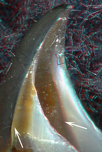
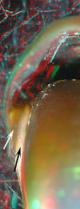
Ancistroteuthis lichtensteini, male, 89 mm ML, 3.0 mm
URL. LEFT - Oblique view. RIGHT - Posterior view.
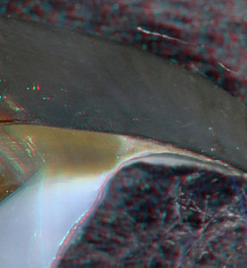
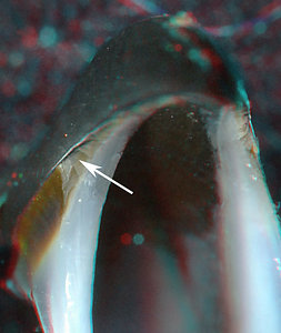
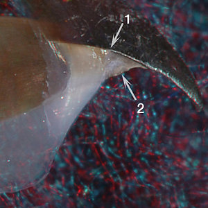
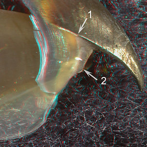
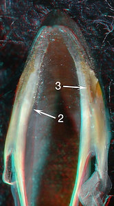
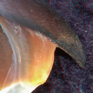
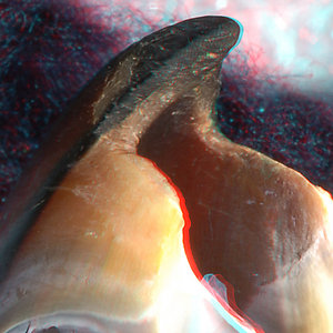
A. lichtensteini, 107 mm ML, 4.8 mm URL, views of shoulder. MIDDLE - Side view. RIGHT -
Front-oblique view.The larger beak (Right) clearly shows a A Step and a Wedge (arrow). The Shoulder Blade of the large beak looks like that of a Bipartite beak (Right) but the smaller beak has a yellowish, dorsal region (Left, left arrow) on/under the Shoulder Blade. A reduced Palatoshoulder Ridge seems to be present (Left, right arrow). The posterior view (Left middle) shows an "outer lobe" (white arrow) and a "middle lobe" (black arrow) that contains the Hyaline Matrix and looks rather similar to lobes 3 and 5 of Ancistrocheirus (see next species). In the enlarged view (click on image), the outer lobe seems to have a groove near the Hyaline Matrix; the middle layer has additional pigment covering it dorsally an slightly medially. This appearance has some similarities to that of Ancistrocheirus seen below.
Ancistroteuthis lichtensteini is an exception in the family. The beaks only indication of a Tripartite shoulder is (1) the yellowish area dorsally on/under the Shoulder Blade in the smaller beak and (2) the presence of a Wedge (Right, arrow) that extends from the Shoulder Blade; the wedge extends onto the Pallet and a step is present between it and the jaw edge which, somewhat, recalls the peculiar anterior portion of the Tripartite Shoulder Blade. Apparently the elongate, slender rostrum and strongly tapering leading edge of the shoulder, which forms an obtuse angle to the Jaw Edge, are responsible for the peculiar shoulder structure. This beak does not qualify as a Tripartite beak at this stage; perhaps more evidence of its Tripartite nature will appear in older beaks.
ANCISTROCHEIRIDAE: Ancistrocheirus lesueurii
UPPER BEAK CHARACTERISTICS
- Step: Present between Palatoshoulder Ridge and Jaw Edge.
- Shoulder Blade: Present on outer-most layer of Shoulder but, in some specimens, an irregularly-shaped, yellow layer seems to lie beneath a translucent Shoulder Blade.
- Shoulder Blade: Consists of one major component with a Wedge located anterior to it.
- Hyaline Matrix: Present within middle layer of Shoulder.
- Palatoshoulder Ridge: Present, large. Extends onto Palate; partially with free-edge in some small beaks.
- Pigmentation of posterior surface of Hyaline Matrix. Partial pigmentation of the middle layer of the Shoulder reveals three lobes with the middle one containing the Hyaline Matrix.
- Yellow Line: Absent.
Phylogenetic relationships. Morphological evidence (e.g., Young and Harman, 1998) at present places the Ancistrocheiridae at the base of the enoploteuthid families; unfortunately, molecular data are lacking.
Ancistrocheiridae Structure
- EXTERNAL FEATURES
Eight beaks have been examined; one beak has been cut.


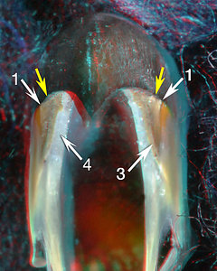
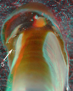
Ancistrocheirus lesueurii, (probable male ?, ML ?), 1.8 mm URL. LEFT - Side view. MIDDLE - Front view. RIGHT - Posterior view.
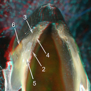
A. lesueurii, (probable female ? , ML ?), 3.4 mm URL. LEFT - Side view. MIDDLE - Front view. RIGHT - Posterior view.
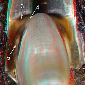
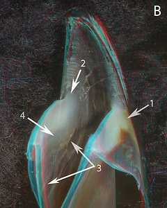
ARROWS: 1 - Posterior end of Jaw Edge and anterodorsal end of Shoulder Blade. 2 - Palatoshoulder Ridge. 3 - Wedge (= lobe 3). 4 - Thick layer lateral to Palatoshoulder Ridge (= lobe 4). 5 - Middle lobe containing the Hyaline Matrix (= lobe 5). 6 - Shoulder Blade. Note the associated yellowish area.
The images of A. lesueurii (above) are of two very different sized beaks that show the same peculiar Shoulder pigmentation. The front views (Middle) show a Wedge (arrow 3) similar to that seen in Ancistroteuthis (previous set of images). The region normally occupied by a thick Hyaline Matrix in Bipartite beaks is partially pigmented in the smallest beaks examined. This region is pigmented anteriorly (Middle images) and dorsally and posteriorly (Right images) in the form of two lobes (3, 4) partially enclosing a ventral, median lobe (5) containing the Hyaline Matrix. In the smaller, 1.8 mm URL beak, the Hyaline Matrix of the of lobe 5 seems to separate the Wedge and the thick layer (4) more at the anterior end than in the larger beak. Also, in the posterior view of the small beak (Top, Right) an unpigmented strip of the Lateral Wall lies medial and posterior to the Shoulder (Top, Right, posterior to 4). This strip is the last area of the Lateral Wall adjacent to the Shoulder to be pigmented and recalls a similar situation in onychoteuthid pigmentation.
The dorsal part of the Shoulder, therefore, is composed of 3 "lobes;" these are more apparent posteriorly than anteriorly. Viewed posteriorly (Right images), the Shoulder has an outer lobe, lobe 3, which seems to be continuous anteriorly with the Wedge; a middle lobe, lobe 5, containing the remaining Hyaline Matrix, which is recognizable in both anterior and posterior views by the Hyaline Matrix; and an inner lobe, lobe 4, that looks almost like a thickened Palatoshoulder Ridge in the anterior view. The three areas seen in the front and posterior views are not always recognizable in fully pigmented shoulders.
- INTERNAL STRUCTURE: Longitudinal cuts through the Shoulder of A. lesueurii, 3.2 mm URL.
- INTERNAL STRUCTURE: Cross-sectional cuts through the Shoulder of A. lesueurii, 3.2 mm URL.
 Click on an image to view larger version & data in a new window
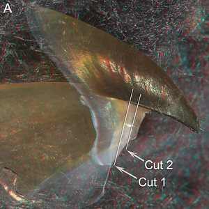
Figure. Side view of the right beak fragment showing where two cross-sectional cuts were made.
- - - - - CROSS-SECTIONAL CUT 1 - - - - -
Click on an image to view larger version & data in a new window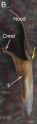

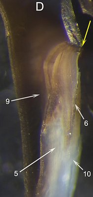
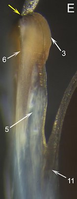
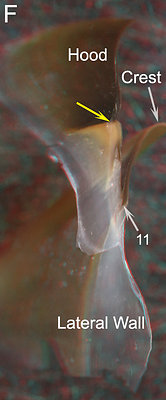
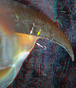
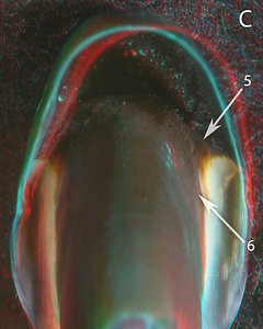
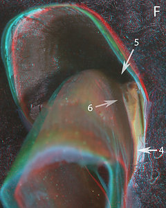
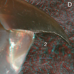
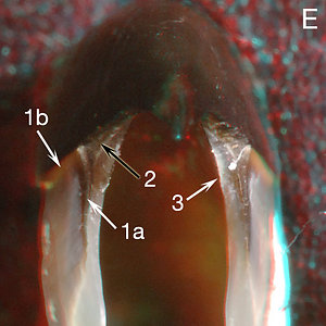
Figure. B - Posterior view. C - Posterodorsal view. D - Photomicrograph (2D), anterior beak fragment, posterior view taken perpendicular to cut surface. E - Photomicrograph (2D), posterior beak fragment, anterior view taken perpendicular to cut surface. F - Anterolateral view of posterior beak fragment to aid viewer orientation.
ARROWS: 3 - Wedge (= lobe 3). 5 - Middle lobe containing Hyaline Matrix (= lobe 5). 6 - Shoulder Blade. 9 - Groove where Free and Sessile Shoulders join. 10 - Edge of cut surface. 11 - Ventral edge of the Palatoshoulder Ridge. Yellow arrows - Point to same spot on all beaks.This cut missed most of the Fused Shoulder. The cut does show the structure of the Free Shoulder including the Shoulder Blade (6), the Wedge (3), and lobe 5 (containing most of the Hyaline Matrix) which lies mostly in the Free Shoulder. The Wedge is a thick pigmented area that lies under most of the thin Shoulder Blade. The growth lines of the Wedge are concave relative to the lateral surface of the Shoulder and accentuate the size of the Step. Much of the structure surrounding lobe 5 is difficult to interpret. Laterally it appears to be bound by a ventral extension of the Wedge that further on dissipates into the Hyaline Matrix (Figs. D, especially E).
- - - - - CROSS-SECTIONAL CUT 2 - - - - -
Click on an image to view larger version & data in a new window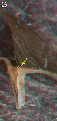
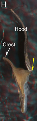
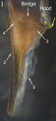

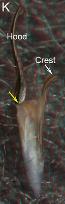
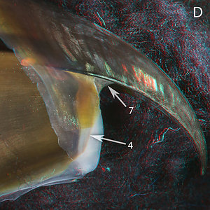
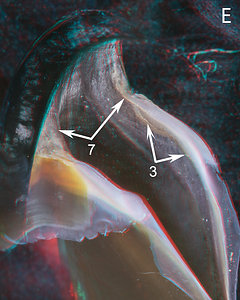
Figure. G - Posterolateral view. H. Posterior view. I - Photomicrograph (2D), anterior beak fragment, posterior view taken perpendicular to cut surface. J - Photomicrograph (2D), posterior beak fragment, anterior view taken perpendicular to cut surface. K - Posterior beak fragment, Posterior view to aid viewer orientation.
ARROWS: 2 - Palatoshoulder Ridge. 3 - Wedge (= lobe 3). 4 - Thick layer lateral to Palatoshoulder Ridge (= lobe 4). 5 - Middle lobe containing Hyaline Matrix (= lobe 5). 7 - Bridge. Yellow arrows - Point to same spot on all beaks.This cut passes through the broad fusion of the Wedge (= lobe 3) and lobe 4; together these lobes extend from the Hood to the Crest. The lobe 3 attachment to the Hood is abrupt while the lobe 4 attachment to the Crest is more graded in pigmentation. A shallow groove on the dorsal surface of the lobes (groove corresponds with arrow 9 of Cut 1) and the poorly seen growth lines, that approximately parallel the dorsal surface, distinguish the two fused lobes.
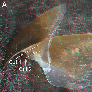
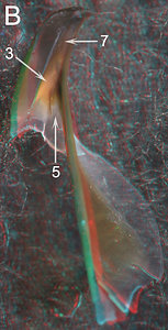
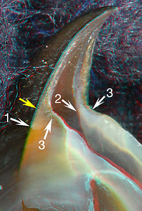
Figure. A - Lateral view of the left half of the beak showing where two longitudinal cuts were made. B - Dorsal-posterior view of the same beak fragment showing a different perspective of the Shoulder and Bridge.
ARROWS: 3 - Wedge or outer lobe (= lobe 3). 5 - Middle lobe containing the Hyaline Matrix (= lobe 5). 7 - Bridge.
- - - - - LONGITUDINAL CUT 1 - - - - -
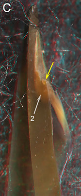
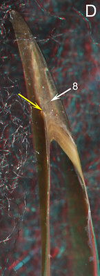
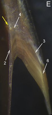

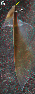
Figure. C - Oral-medial view. D - Oral (=ventral) view. E - Photomicrograph (2D), dorsal beak fragment, oral view taken perpendicular to the cut surface. F - Photomicrograph (2D), ventral beak fragment, aboral (= dorsal) view taken perpendicular to cut surface. This is the other beak surface from the cut. G - Aboral view with slight medial tilt of the ventral beak fragment; aids viewer orientation of Fig. F.
ARROWS: 2 - Palatoshoulder Ridge. 3 - Wedge (= lobe 3, posterior view). 4 - Thick layer lateral to Palatoshoulder Ridge (= lobe 4). 6 - Shoulder Blade. 8 - Dark band apparently continuous with the central mass of the Shoulder. Yellow arrows - Point to same spot on all beaks.
Cut 1 shows a well-pigmented Shoulder (it missed the middle lobe (5) and its Hyaline Matrix - see Fig. H below) but a small amount of irregularly-shaped hyaline material is present on the anterior suface of the Shoulder. The Palatoshoulder Ridge (E, 2, F, 2) is much more darkly pigmented than the main mass of the Shoulder; in the latter growth lines can be seen. The main mass is composed of the Wedge (E, 3) and lobe 4 (see images under "External features") and the two lobes are smoothly continuous and exhibit chevron-shaped growth lines that parallel the posterior surface of the Shoulder (E, F). The two lobes can be distinguished by a light "line" that extends along the apex of the growth lines. Fig. C shows the extent of the Palatoshoulder Ridge (2) via scattered back-lighting from the more lightly pigmented portions of the beak.
- - - - - LONGITUDINAL CUT 2 - - - - -
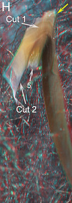
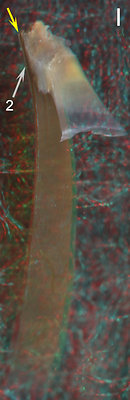
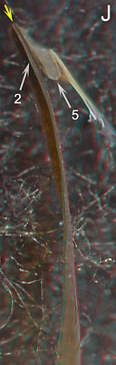

Figure. H - Similar view to Fig. G, with a more posterolateral tilt, taken after Cut 2 was made; the image shows where the two cuts were made on this beak fragment. I - Oral-lateral view of Cut 2. J - Oral (= ventral) view of Cut 2. K - Photomicrograph (2D), oral view taken perpendicular to cut surface.
ARROWS: 2 - Palatoshoulder Ridge. 5 - Middle lobe containing the Hyaline Matrix (= lobe 5). Yellow arrows - Point to same spot on all beaks.
The oblique, aboral angle of Fig. H shows that Cut 2 passed through a portion of the middle lobe (5) that had no pigment on its mid-posterior surface. The oral view of Cut 2 shows the Hyaline Matrix, of lobe 5, extending from the anterior surface of the Shoulder to its posterior surface (Figs. I-K).
Comments.
The beak of Ancistrocheirus is very peculiar. The presence of a lateral Shoulder Blade, a large Palatoshoulder Ridge and a Step exclude it from Tripartite and Unipartite categories; its peculiar pigmentation of the posterior Shoulder excludes it from the Bipartite category. The beak, however, has some major similarities to the Tripartite beak. The Ancistrocheirus Shoulder has a thick, dorsal, pigmented region, formed by lobes 3 and 4, that spans the gap between the Crest and the Hood and corresponds to the dorsal portions of the Inner and Outer Components of the Shoulder Blade in Tripartite beaks. In the latter, these two Components are identified by a dorsal groove that is continuous posteriorly with the groove formed at the junction of the Free and Sessile Shoulders, and the distinctive associated growth lines. This same groove is present in the Ancistrocheirus Shoulder along with the apparently similar growth lines (the lines are not nearly as distinct as those of Onychoteuthis). Presumably in Ancistrocheirus, as in Tripartite beaks, this area of the Shoulder is formed from the Bridge. One other major difference is apparent regarding the position of the Hyaline Matrix. In Tripartite beaks a distinct, complete layer of Hyaline Matrix lies medial to the Shoulder Blade. In Ancistrocheirus the Hyaline Matrix lies in the middle of what would correspond to the center of the Tripartite Shoulder Blade.
One could easily imagine the beak of Ancistrocheirus as an early offshoot of a line leading to the Tripartite Shoulder, although the potential homologous structures are not all obvious due to the unusual position of the Hyaline Matrix and the peculiar posterior pigmentation of the Shoulder.
CYCLOTEUTHIDAE: 2 genera
UPPER BEAK CHARACTERISTICS
- Step: Present. Between thick Wedge and Jaw Edge.
- Shoulder Blade: Present. On outer layer of Shoulder but in some specimens a yellow layer seems to lie beneath the translucent Shoulder Blade.
- Shoulder Blade: Consists of one major component with a Wedge located anterior to it.
- Hyaline Matrix: Forms middle layer of Shoulder.
- Palatoshoulder Ridge: Present. Extends onto Palate; no free-edge, but almost.
- Pigmentation of posterior surface of Hyaline Matrix: Appears to have "leaf-shape" pigmentation.
- Yellow Line: Absent. Yellow Line imposter present in young Cycloteuthis; dorsally located yellow patch present in D. laciniosa.
Phylogenetic relationships. The genus Discoteuthis was originally placed in the Cycloteuthidae primarily due to similarities with Cycloteuthis in the shape and position of the funnel-mantle locking-apparatus. DNA studies by Lindgren (2010) strongly supported this relationship (all three analyses place the two genera together with good to strong support) while that of Lindgren, et al.(2012) placed the two genera far apart but with weak support. We accept the results of Lindgren (2010) as the more likely since they agree with the morphology.
At the family level, there is no good morphological evidence for the placement of the Cycloteuthidae. In the 2010 molecular study Lindgren placed the Cycloteuthidae as a sister taxon to the Brachioteuthidae in her Bayesian (with strong support) and Likelihood (with weak support) analyses but to the Mastigoteuthidae in her parsimony (with weak support) analysis. The 2012 study, which didn't recognize Cycloteuthidae with two genera, placed Discoteuthis as a sister taxa (with weak support) to a clade containing the lepidoteuthid and chiroteuthid families. However, Cycloteuthis was placed as a sister taxon (with weak support) to a clade containing the Brachioteuthidae, Gonatidae, Pyroteuthidae, Enoploteuthidae and Onychoteuthidae. Assuming the Cycloteuthidae, with two genera, is a natural group, as indicated in the first paragraph, and that in the 2012 study the position of Cycloteuthis (rather than Discoteuthis) represents the phylogenetic position of the family, we are left with some intriguing possibilities. See "Comments" below. (This tree diagram is seen under Onychoteuthidae, Tripartite Beaks.)
Cycloteuthidae: External features
We examined three beaks of Cycloteuthis sirventi, two beaks of Discoteuthis discus and one beak of D. lacinosa. We have relied on D. laciniosa to represent this family since it is the only species for which we have a critical stage. This beak, unfortunately, was not available to cut.
 |  |  |  |
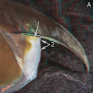
| Discoteuthis laciniosa, immature female, 128 mm ML, 5.8 mm URL. LEFT - Side view. LEFT-MIDDLE - Oblique view. RIGHT-MIDDLE - Oral view. RIGHT - Oral-front view. | |||
 |  |  |
| D. laciniosa, same beak as above. LEFT - Front view. MIDDLE - Posterior view. | D. laciniosa, immature male, 52 mm ML, 2.0 mm URL. RIGHT - Posterior view. | |
ARROWS: 1 - Posterior end of Jaw Edge and anterodorsal end of Shoulder Blade. 2 - Palatoshoulder Ridge. 3 - Large Wedge. 4 - Hyaline Matrix. 5 - Posterior junction between Free and Sessile Shoulder. Yellow arrow - Step.
The Wedge (3) in D. laciniosa (above) is unusual in being very large and thick, appearing much like the "Shoulder Blade" of Tripartite beaks, especially that of onychoteuthids. The crease (5) in the posterior pigmentation of the young D. laciniosa recalls the leaf-shaped pigmentation in young Tripartite beaks with a crease that lies at the posterior junction of the free and fused parts of the shoulder. A distinct Palatoshoulder Ridge is present (2) but has not covered much of the Shoulder at this size. With the exception of the distinct lateral Shoulder Blade, this beaks looks very much like a Tripartite beak. However, only cuts through the Shoulder of D. laciniosa beaks will reveal how similar it is to Tripartite beaks.
The largest D. discus we have seen (107 mm ML, 6.1 mm URL) has only the slightest indication of a Wedge and Step, while in small D. laciniosa (52 mm ML, 2.0 mm URL) with far less pigment in the Lateral Wall than its larger congener, a distinct Wedge and Step is present. Cycloteuthis sirventyi at 200 mm ML (6.0 mm URL) has a very small wedge and step. The Lateral Wall that is posterior to the Shoulder, however, is almost entirely unpigmented at this size. In a mature female (375 mm ML, 12.8 mm URL; the next largest beak we have seen) there is a slight Step but little other evidence remains of the structure of the Shoulder. As a result, we have seen no beaks of Cycloteuthis that are close to a critical stage in this species.
Comments
The considerable differences in the beaks of the three genera of the Cycloteuthidae is surprising and requires a more detailed look. It was also unexpected that the beak of Discoteuthis laciniosa would have so much in common with Tripartite beaks (i.e, onychoteuthids, enoploteuthids, lycoteuthids). However, the Lindgren, et. al. (2012) study found the Cycloteuthidae (as considered above) as a possible sister group to a clade containing the Brachioteuthidae, Gonatidae, Pyroteuthidae, Enoploteuthidae and Onychoteuthidae. Here are two of the Tripartite-beak families; we argued elsewhere (Pyroteuthidae, under 3° Unipartite Beaks) that the Pyroteuthidae may have secondarily lost the Tripartite features. While the 2012 phylogeny if these taxa may not be entirely correct, it at least suggests the possibility that the Cycloteuthidae may be in the lineage that lead to the full development of the Tripartite beak. Of course, there may have been several independent attempts at the evolution of the Tripartite beak. In either case a better understanding of the structure of the cycloteuthid beaks and those of the Ancistrocheiridae may provide useful information on how Tripartite beaks evolve.
Lepidoteuthid families 1: OCTOPOTEUTHIDAE & LEPIDOTEUTHIDAE: 3 genera
UPPER BEAK CHARACTERISTICS
- Step: Present. Between Hyaline Matrix (or its pigmented replacement in an older beak) and Jaw Edge.
- Shoulder Blade: Present on outer-most layer of Shoulder; small.
- Shoulder Blade: Consists of one pigmented component.
- Hyaline Matrix: Shoulder virtually all Hyaline Matrix except for small Shoulder Blade and except in very large beaks. In very large beaks, Outer Part and posterior surface of Shoulder become pigmented; Hyaline Matrix withdraws from Inner Part but remains in Middle Part of Shoulder.
- Palatoshoulder Ridge: Absent.
- Pigmentation of posterior surface of Hyaline Matrix: Not well known. Appears to begin dorsally and, perhaps, medially.
- Yellow Line: Absent.
Phylogenetic relationships. These two families have been placed together in the Tree of Life, along with Pholidoteuthidae in the "Lepidoteuthid families", on the basis of morphology. The support for the relationship between Lepidoteuthidae and Octopoteuthidae is much stronger than the support for all three, so Pholidoteuthidae is treated separatedly below (see Lepidoteuthid families 2: Pholidoteuthidae)). One of the major morphological characters relating the two families was similarity of the beaks. Lindgren (2010), on the basis of molecular characters, placed the two families together, in all three analyses with with weak to excellent support. Lindgren, et al., 2012 also placed them together (with good support).
Octopoteuthidae & Lepidoteuthidae: Structure
- EXTERNAL FEATURES
We examine the beak of Octopoteuthis which should represent the beak of Lepidoteuthis fairly well. The beak of Taningia appears be somewhat different (in spite of its closer relationship to Octopoteuthis) but we have no critical-stage beaks to cut.
Octopoteuthis danae, sex ?, 155 mm ML, 8.9 mm URL. LEFT - Side view. MIDDLE - Oblique view. RIGHT - Posterior view.
Octopoteuthis danae, sex ?, 205 mm ML, 11.2 mm URL. LEFT - Side view. MIDDLE - Oblique view. RIGHT - Posterior-oblique view ARROWS: 1 - Shoulder Blade. 2 - Anterior ridge of Hyaline Matrix that extends onto Palate. 3 - Thickening of the Lateral Wall that marks approximate anterior and dorsal ends of Hyaline Matrix. 4 - Anterior end of pigmented outer layer of Lateral Wall seen through Hyaline Matrix. 5 - Bridge region. 6 - Lateral curvature of Lateral Wall. 7 - Broad, pigmented ridge replaces ridge 2.
In the smaller beak (A-C) the Shoulder Blade (A, 1; B, 1) is narrow but distinct; new pigment of the Outer Part of the Shoulder is present mostly ventral to the Shoulder Blade but apparently continuous with it posteriorly. The entire anterior edge of the Shoulder is composed of Hyaline Matrix which extends medially and posteriorly to a pigmented thickening of the Lateral Wall (B, 3) and laterally and posteriorly to just cover the anterior edge of a lateral bend of the Lateral Wall (B, 4). The embayment lateral to the anterodorsal edge of the Hyaline Matrix (marked by the anterior edge of the Shoulder Blade (A, B)) creates a small Step between the Hyaline Matrix and the Jaw edge. In the larger beak (D-F), this anterior portion of the Hyaline Matrix is replaced by a pigmented ridge (D, 7, E, 7) which is continuous with the pigmented Lateral Wall. The Shoulder Blade is barely recognizable (D) as the pigment of the Outer Part of the Shoulder has greatly increased. The Step, however, is more recognizable. The medial and lateral posterior extent of the Hyaline Matrix is greatly reduced (D, 4; E, 3) and the lateral bend of the Lateral wall (F, 6) is more apparent.
- INTERNAL STRUCTURE: Longitudinal cuts through the Shoulder
A. Large beak, Octopoteuthis danae, 155mm ML, 8.9 mm URL.
Click on an image to view larger version & data in a new window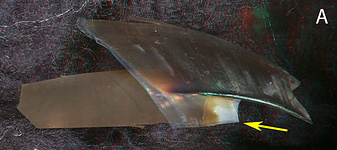
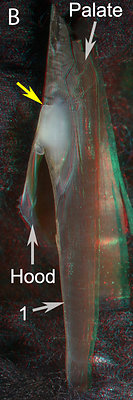

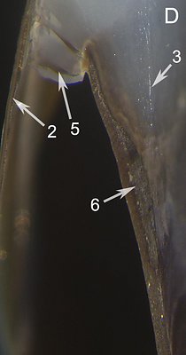

Figure. Octopoteuthis 155mm ML, 8.9 mm URL. A - Side view of beak fragment that was cut longitudinally through the Shoulder. B - Oral view of the Shoulder cut and its beak fragment. C - Same view, higher magnification of cut Shoulder. D - Photomicrograph (2D), oral view taken perpendicular to the cut. E - Oral view of cut Shoulder with the posterior end tilted toward the viewer.
ARROWS: 1 - Lateral Wall. 2 - Pigment of the Outer Part of the Shoulder. 3 - Medial edge of the Hyaline Matrix in the plane of the cut. Lower arrow indicates the posterior limit of the Hyaline Matrix. 4 - Lateral bend of the Lateral Wall. 5 - Crack in the Hyaline Matrix where the Matrix lies between the anterior edge of the Lateral Wall and the pigmented Outer Part of the Shoulder. The crack (and the one above it) probably are artifacts from the cutting process. 6 - Thickened area of the Lateral Wall. Yellow arrows - Point to the same spot on all beaks to aid viewer orientation.The lateral bend in the Lateral Wall (C, 4) places this pigmented layer over part of the posterior surface of the Hyaline Matrix (C, 3). This anterior edge of the Lateral Wall (C, D), however, lies within the Hyaline Matrix (D). Pigment of this tip impings laterally into the Hyaline Matrix (D use magnified image). This latter pigment (E, 7) incorrectly appears in to be on the posterior surface of the Hyaline Matrix but an irregular layer of Hyaline Matrix covers this pigment posteriorly but it is too transparent to be seen in this photograph.
- INTERNAL STRUCTURE: Cross-sectional cut through the Lateral Wall.
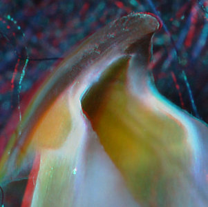
A. Larger beak, Octopoteuthis danae, 205mm ML, 11.2 mm URL: *** Cut 3 ***
Click on an image to view larger version & data in a new window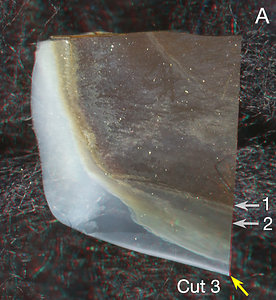
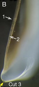
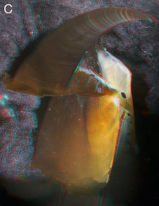
Figure. A - Medial view of the anteroventral corner of the Lateral Wall (the mirror image of this portion in Fig. A of the Shoulder cuts). The anterior end of the beak is at the left of the image where the thick, opaque white Hyaline Matrix is. B - Photomicrograph (2D), anterior view taken perpendicular to the cut surface. C - Lepidoteuthis grimaldii, 22 mm URL, from sperm whale stomach. Oblique view with the anterior end tilted toward viewer.
ARROWS - 1 - Arrow indicates a very thin layer of hyaline material. 2 - A thicker layer of Hyaline Matrix.
Cut 3 was made to determine the extent of Hyaline Matrix along the ventral region of the beak compared to an extreme case in Lepidoteuthis. In the ventral part of the cut (2), the Hyaline Matrix is thicker than the pigmented layer of the Lateral Wall. Compare this with the Hyaline Matrix of a large Lepidoteuthis (C) taken from a sperm whale stomach: On the left side of the beak the Hyaline Matrix is white (presumably an artifact of whale digestion); on the right side the Hyaline Matrix is detectable as the yellow light penetrating the Lateral Wall pigment. The much thicker and more extensive Hyaline Matrix of the Lepidoteuthis beak provides an important difference to a beak which is otherwise very similiar an Octopoteuthis beak.
- Step: Present. However very small and between Wedge and Jaw Edge.
- Shoulder Blade: Present on Outer Part of Shoulder but with a white/translucent thin layer over its lateral (= outer) surface.
- Shoulder Blade: Consists, apparently, of one major component with a Wedge located anterior to it.
- Hyaline Matrix: Forms middle layer of Shoulder.
- Palatoshoulder Ridge: Present. With free-edge extending onto Palate in larger beaks of P. adami.
- Pigmentation of posterior surface of Hyaline Matrix: Not understood. Large P. adami with indications of an outer and middle lobe in posterior view.
- Yellow Line: Often present but weak and diffuse ventrally; structure unknown; appearance more like that of an onychoteuthid than an ommastrephid.
- Step: Absent.
- Shoulder Blade: Shoulder Blade thick; mostly covered laterally by thin layer of hyaline-like material.
- Shoulder Blade: Consists of two components (Inner and Outer). Components separated by distinct line.
- Hyaline Matrix: Apparently forms very thin middle layer of Shoulder. Layer lightly pigmented and tends to blend with Palatoshoulder Ridge medially and Inner Component of Shoulder Blade laterally.
- Palatoshoulder Ridge: Present. Without sharply defined edges and without free edges.
- Yellow Line: Absent.
- EXTERNAL FEATURES
We have seen only 1 beaks from each of two species in this very large family. The larger beak (Sepia latimanus, 4.1 mm URL) is well pigmented and husky. Only the smaller beak (Sepia sp., 1.0 mm URL) was at a critical state of development and was cut.
Sepia sp., 1.0 mm URL. LEFT - Oblique view.
Sepia latimanus, 145 mm ML, 4.1 mm URL. MIDDLE - Side view. RIGHT - Oblique view.
The Left image shows the Shoulder Blade, the Palatoshoulder Ridge and a hazy white layer between them which is probably the Hyaline Matrix. The Middle image shows the absence of a Step. All three images show the absence of a Yellow Line.
- INTERNAL STRUCTURE: Longitudinal cut through the Shoulder. Click on an image to view larger version & data in a new window
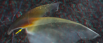
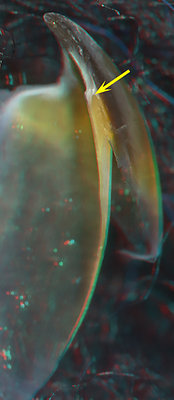
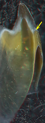

Figure. Sepia sp. 1.0 mm URL. TOP: Side view showing the position of the cut (arrow). BOTTOM: Left - Oral-lateral view of cut beak. Arrow points to anterior end of the Shouder Blade. Middle - Oral view of cut beak. Right - Photomicrograph (2D), oral view taken perpendicular to the cut beak.
ARROWS: 1 - Line between the two parts of the Shoulder Blade. 2 -Hyaline Matrix (?). 3 - Palatoshoulder Ridge. Yellow arrows - Point to the same spot on all beaks to aid viewer orientation.The cut through the shoulder shows that the thick Shoulder Blade occupies most of the Shoulder. A peculiar middle layer (2) exists between the Palatoshoulder Ridge (3) and the Inner Component of the shoulder Blade. We assume this partially pigmented layer is a reduced Hyaline Matrix that, strangely, seems to blend into the structures on either side. A thick layer of hyaline material covers the anterior end of the Shoulder Blade and a thin layer covers the lateral surface of the Shoulder Blade (not visible in the photograph).
Comments
The double shoulder blade and absence of a Step is suggestive of a Unipartite beak but the presence of a large Palatoshoulder Ridge is suggestive of a Bipartite beak. The near absence of the Hyaline Matrix and the indistinct appearance of the shoulder structures makes it more peculiar. Whether or not this beak will be characteristic of the family remains to be seen.
B. Larger beak, Octopoteuthis danae, 205mm ML, 11.2 mm URL.
We have made two longitudinal cuts through the Shoulder of this beak and one cross-sectional cut through the Lateral Wall.
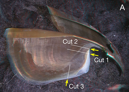
Figure. A - Medial view of the left half of the cut beak showing approximately where the "structural" cuts were made on the right half of the beak.
***** CUT 1 *****
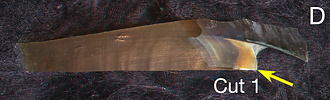
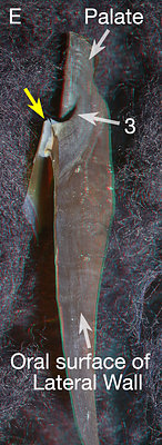
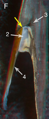
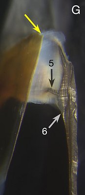
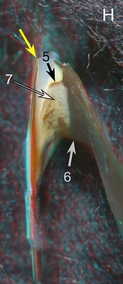
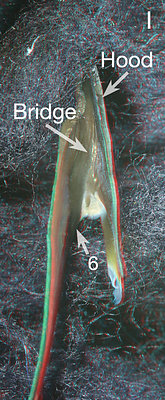
Figure. Octopoteuthis 205mm ML, 11.2 mm URL. D - Side view of beak fragment showing where cut was made. E - Oral view of the same beak fragment. F - Closer oral view of cut surface of the Shoulder. G - Photomicrograph (2D), oral view taken perpendicular to the cut surface. H - Oral(= ventral)-posterior view; the cut beak is tilted to show more of the posterior surface of the Shoulder. I - Aboral(= dorsal)-posterior view of the same fragment
ARROWS: 2 - Hyaline Matrix. 3 - Rounded, pigmented ridge onto Palate. 4 - The Free Shoulder. 5 - Pigmented "finger" extending into Hyaline Matrix. 6 - Lateral bend of the Lateral Wall. 7 - Pigmented plate partially covered by Hyaline Matrix which is continuous with the pigmented "finger" (5). Yellow arrows - Point to the same spot on all beaks to aid viewer orientation.
The pigmentation of the Shoulder has changed considerably in this larger beak. The Hyaline Matrix has disappeared almost entirely from the medial surface of the Shoulder and the small remaining bit has been undercut by extension of the Lateral Wall (anterior "finger" in F) suggesting that this Hyaline Matrix would not have remained for long. The two heavily pigmented "fingers" seen extending laterally from the Lateral Wall in the cut are edges of pigment sheets; the anterior sheet apparently is destined to cover much or all of the near-anterior end of the Hyaline Matrix and the posterior one the near-posterior end. Fig. I shows how the lateral bend of the Lateral Wall (6) seems to be continuous with the Bridge and therefore a LW-P Contiuum. However, in (G, 6) much of this bend seemingly results from a thickening of the Lateral Wall.
***** CUT 2 *****
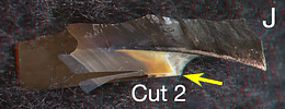
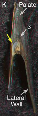
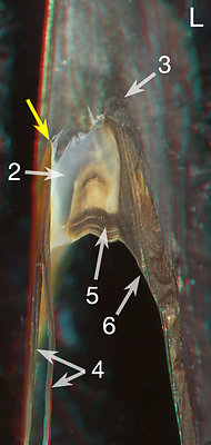
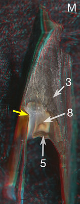
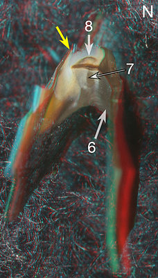
Figure. J - Side view of beak fragment showing where cut was made. K - Oral view of the same beak fragment. L - Closer oral view of cut surface with greater resolution. M - Oral-anterior view; the Palate is tilted slightly toward the viewer. N - Oral-posterior view; the Palate is tilted strongly away from the viewer.
ARROWS: 3 - Rounded, pigmented ridge onto Palate. 4 - Free Shoulder broken due to cut. 5 - Pigmented "finger" extending into Hyaline Matrix. 6 - Lateral bend of the Lateral Wall. 7 - Pigmented plate partially covered by Hyaline Matrix which is continuous with the pigmented "finger" (5). 8 - Internal pigmented ridge (see M, N). Yellow arrows - Point to the same spot on all beaks to aid viewer orientation.
This more anterior cut through the Shoulder simply shows a more advanced state of pigmentation of the Hyaline Matrix and how complex this pigmentation process is.
Comments
The octopoteuthid beak has a very unusual shape: (1) The Lateral Wall, in profile, almost forms a rectangle beneath the hood and with the Shoulder forming the entire anterior end of the rectangle; (2) The Rostrum is very long and very recurved. It is similar to Bipartite beaks in having a simple (i.e., one component) Shoulder Blade but differs in the absence of a Palatoshoulder Ridge and the presence of an insertion by the Lateral Wall into the middle of the Hyaline Matrix (see D & E in the 8.9 mm URL beak). The lateral bend of the Lateral wall at the Shoulder appears to be continuous with the Bridge and appears to be a form of the LW-B Continuum. The full pigmentation of the Hyaline Matrix is complicated and not well understood. The beak has a Step but not a conventional one.
Lepidoteuthid families 2: PHOLIDOTEUTHIDAE: 1 genus
UPPER BEAK CHARACTERISTICS
Phylogenetic relationships. We place this family within the "lepidoteuthid families" (Pholidoteuthidae, Lepidoteuthidae and Octopoteuthidae) on the basis of morphology and especially the similarity of the dermal cushions shared by Pholidoteuthis and Lepidoteuthis. The position of Pholidoteuthis within the lepidoteuthid families, however, is not well secured by morphology. The DNA analysis of Decapodiform relationships by Lindgren (2010) and Lindgren et al. (2012) shows that the Pholidoteuthidae falls either within the lepidoteuthid or chiroteuthid families or between them but generally without good support in either position.
Pholidoteuthidae: External features
Pholidoteuthis contains two species. No beaks were available for cutting.
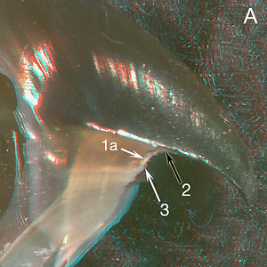 |  |  |
| Pholidoteuthis adami, 281 mm ML, 4.3 mm URL. A - Side view. B - Front view. C - Posterior view. | ||
 |  | 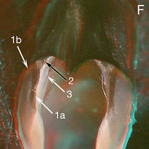 |
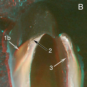
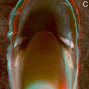
| Pholidoteuthis massyae, male, 122 mm ML, 2.9 mm URL. E - Side view. F - Front view. | P. massyae, male, 161 mm ML, 3.6 mm URL. E - Front view. | |
ARROWS: 1a - Anterior edge of Shoulder Blade. 1b - Light area beneath Shoulder Blade. 2 - Wedge. 3 - Palatoshoulder Ridge.
Both species of Pholidoteuthis, on first look, appear to have a typical Bipartite Type 1 beak due to its large Palatoshoulder Ridge and apparent lack of a Step. Surprisingly, P. adami has an oblique Jaw Angle, and P. massyae has an acute Jaw angle. However, a Step between the Jaw Edge and a small Wedge is present but very small. The "Wedge" is difficult to recognize (arrow 2). It is a thickened region arising beneath the anterodorsal end of the Shoulder Blade and extending onto the Palate where it quickly becomes indistinguishable. The beak often has a yellowish line (E, 1b, F, 1b) or sometimes a patch (B, 1b) apparently lying beneath the dorsal region of the thin Shoulder Blade (E, F). What appears to be a thick Shoulder Blade (E) may be part of the Wedge. In the P. massyae beak there appears to be a white Outer Covering (F) over part of the Shoulder Blade. In a posterior view of the P. adami beak there are, at least, two distinct lobes with the medial one containing the Hyaline Matrix (we have not seen a posterior view of a P. massyae beak at a critical stage).
Comments
The upper beak of Pholidoteuthis is atypical but we have insufficient information to understand its structure. However, it seems to have some similarities to the onychoteuthid Tripartite beak (i.e., Outer Cover (not always present), Wedge, two lobes in the posterior view of the Shoulder).
ORDER SEPIOIDEA, Suborder:Sepiida
SEPIIDAE - 3 genera, >100 species
UPPER BEAK CHARACTERISTICS
Phylogenetic relationships. The Sepiidae is a very large and distinctive family. In the Tree of Life it is placed in the Sepioidea along with the Sepiolida. The Sepiidae, Sepioida, Myopsida, Spirulida and Idiosepiidae are thought to be near the base of the Decapodiformes, but the relationships among these five taxa is unresolved (Lindgren et al., 2012).
Sepiidae: Structure
COMMENTS ON ATYPICAL BEAKS
Seven taxa are placed in this category. Three taxa (Octopoteutidae/Ledidoteuthidae, Sepiidae) have two distinct types of beaks that show no clear evidence of being more closely related to any one of the three major beak categories. Of the remaining 4 taxa one (Ancistroteuthis) is an unquestioned member of the Onychoteuthidae whose other members have Tripartite beaks. Ancistroteuthis presumably lost most Tripartite features in the process of developing a beak with a very un-onychoteuthid shape. But some of its beak features have (especially, a Wedge) shown up in the beaks of the three other Atypical taxa. Of these, two other taxa, Discoteuthis laciniosa (Cycloteuthidae) and Ancistrocheirus lesueurii (Ancistrocheiridae) have beaks that show strong similarities to Tripartite beaks. Some morphological evidence places Ancistrocheiridae close to the Tripartite taxa so the similarity there may not be surprising. It is surprising, however, that Discoteuthis laciniosa beaks may be even more similar to Tripartite beaks yet the other two species in the family have quite different beaks; the phylogenetic relationships of this family are not resolved. The beaks of the remaining taxa (Pholidoteuthidae), also shows some indication of Tripartite affinities but this family appears to be most closely related to Octopoteuthidae+Lepidoteuthidae.
Unfortunately of the four taxa whose beaks show similarity to Tripartite beaks, only Ancistrocheirus lesueurii has a structure that has been studied by making sections through the beak. Clearly a more intensive study of the beaks of the other three families may help considerably in understanding phylogenetic relationships as well as understanding how Tripartite beaks evolve whether phylogeneticly related or not.


