

Figure. Stoloteuthis sp. A, female, 12 mm ML. Left - Ventrolateral view. Right - Ventral view with mantle cavity opened. Photographs by R. Young.
- Arms
- Suckers globular with small aperatures; suckers approximately equal in size between arms; suckers with circularis muscles.
- Deep web joins all arms to each other except arms IV.
- Suckers globular with small aperatures; suckers approximately equal in size between arms; suckers with circularis muscles.
- Tentacles
- Tentacular club short, coiled aborally. Suckers in numerous irregular series.Tip of club without suckers. Suckers increase in size distally; largest suckers about 0.04 mm in diameter.
- Dorsal region of oral surface of club, immediately distal to tentacular organ, with series of folds.
- Tentacular organ present; barely overlaps base of sucker-bearing club.
- Tentacular stalk broader than club.
- Tentacles completely retractable into pockets via coiling.
Click on an image to view larger version & data in a new window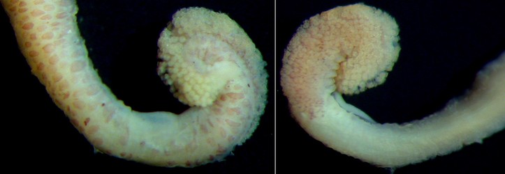
Figure. Tentacular club of Stoloteuthis sp. A, female, 12 mm ML. Left - Aboral view. Right - Oral view. The tentacle organ is visible in both views. Photographs by R. Young.
Click on an image to view larger version & data in a new window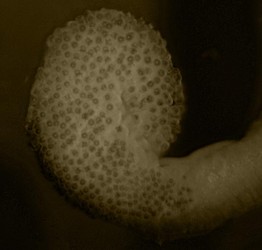
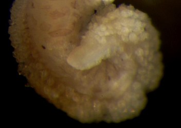
Figure. Tentacular club of Stoloteuthis sp. A, female, 12 mm ML. Left - Oral view. Note distal increase in sucker size. Right - Oral view of club tip. Note that the tip lacks suckers. Photographs by R. Young.
- Tentacular club short, coiled aborally. Suckers in numerous irregular series.Tip of club without suckers. Suckers increase in size distally; largest suckers about 0.04 mm in diameter.
- Head
- Eye with cornea; secondary eyelid at anteroventral half of eye.
- Olfactory organ not recognizable.
- Funnel
- Funnel valve could not be found.
- Dorsal funnel organ poorly defined but small anterior tip and slight medial ridge present. Ventral organs not detected except for a possible anterior lobe of right organ.
- Fins
- Fins short; fin length about half ventral mantle length.
- Fins attach on posterior half of mantle.
- Posterior margin of fins rounded.
- Pigmentation
- Funnel with distinctive pigment pattern: Bare region midventral at bend; pair of bare regions just posterior from this; single line of chromatophores extends to each funnel locking-apparatus and beneath it. Dorsal surface of funnel unpigmented except at tip.
Click on an image to view larger version & data in a new window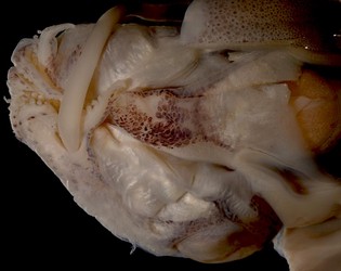
Figure. Ventral view of funnel of Stoloteuthis sp. A, female, 12 mm ML, showing chromatophore pattern. Photograph by R. Young.
- Fins with scattered chromatophores dorsally, none ventrally.
- Thick, silvery patch of iridophores on posteroventral surface of funnel, overlaps most posterior row of funnel chromatophores and lies over anterior region of ventral shield (see photograph above and labeled photograph under "Photophores").
- Clear patch of tissue ventral to eye and anterior to lateral funnel adductor apparently absent.
- Aboral surface of tentacular stalk and club covered with numerous, non-overlapping chromatophores. Oral surface of club with many, small chromatophores beneath suckers.
- Lining of mantle cavity (mantle and visceral cover) without chromatophores.
Click on an image to view larger version & data in a new window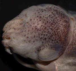
Figure. Anterodorsal view of the head and arms of Stoloteuthis sp. A, female, 12 mm ML, showing chromatophore pattern. Photograph by R. Young.
- Funnel with distinctive pigment pattern: Bare region midventral at bend; pair of bare regions just posterior from this; single line of chromatophores extends to each funnel locking-apparatus and beneath it. Dorsal surface of funnel unpigmented except at tip.
- Photophores
- Pores to visceral photophore seem to be more lateral than typical in Stoloteuthis but ducts penetrate directly through "lens" into photophore.
Click on an image to view larger version & data in a new window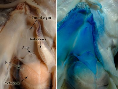
Figure. Ventral view of the internal surface of the funnel and the visceral photophore of Stoloteuthis sp. A, female, 12 mm ML. Left - Unstained. Right - Stained with methylene blue stain. Photographs by R. Young.
- Pores to visceral photophore seem to be more lateral than typical in Stoloteuthis but ducts penetrate directly through "lens" into photophore.
- Viscera: Females
- Nidamental glands large (each 2.5 mm x 1.3 mm) but not as large as expected if animal mature.
- Oviducal opening with two long, slender flaps.
Click on an image to view larger version & data in a new window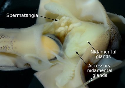
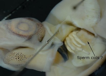
Figure. Mated female Stoloteuthis sp. A, 13 mm ML. Left - Ventral view of opened mantle cavity showing the attachment of large spermatangia. Right - Ventrolateral view of the same specimen with gill cut and funnel retractor pulled forward to show depth of attachment of spermatangia and within the visceral envelope what appears to be sperm coils, presumably the coiled slender ends of the spermatangia.
- Viscera: Males
Click on an image to view larger version & data in a new window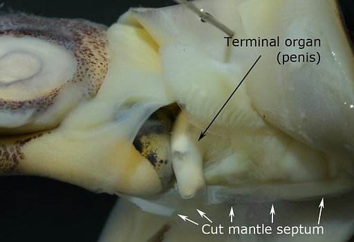
Figure. Ventrolateral view of Stoloteuthis sp. A, male, 16 mm ML showing the terminal organ. Note, also, the ventral mantle septum cut during dissection showing that it is continuous without a broad posterior opening like that found in Heteroteuthis spp..
- Measurements of the, immature 12 mm ML female: Ventral mantle lenght - 17 mm; shield length - 7 mm; dorsal mantle length - 12.3 mm; mantle width - about 11 mm; club length - 2.5 mm; largest club suckers - 0.04 mm in diameter; fin length - 8.9 mm; fin width - 5.7 mm; arm I length - 5.5 mm; arm II length - 5.4 mm; arm III length - 6.2 mm; arm IV length - 5.6 mm; largest arm suckers - 0.3 mm in diameter.


