
Figure. Ventral views of the integumental photophores. Left - Photograph of the preserved squid. Middle - Outline drawing from photograph with all integumental photophores represented by colored dots. Right - Photograph with colored dots superimposed on integumental photophores. Red dots - Complex photophores. Blue dots - Non-complex photophores. Images by R. Young.
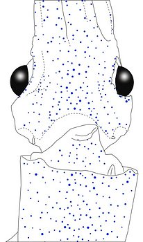

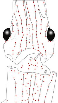
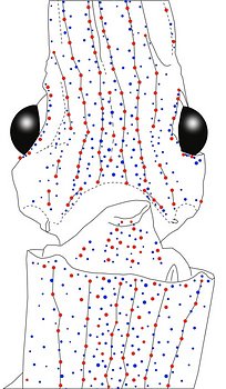
Figure. Same squid, same view as above. Left - Drawing showing only the non-complex (blue) photophores. Left middle - Drawing showing only the complex (red) photophores. Right middle - Same as previous drawing with lines connecting red photophores to aid comparisons of photophore patterns between species. Right - Drawing showing all photophores and lines showing red photophore patterns. Images by R. Young.
spnc4m19obliqpictsm.400a.jpg)
spnc4m19obliqphotsm1.400a.jpg)
spnc4m19obliqdrawrbpattsm1.400a.jpg)
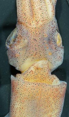
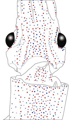
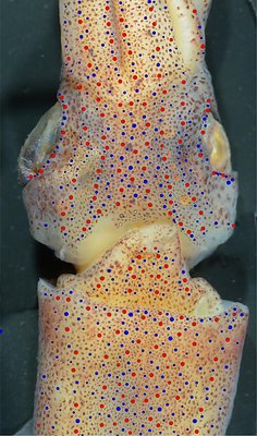
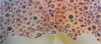
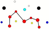
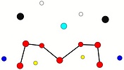
Figure. Ventrolateral view of the head and proximal regions of the arms of the same squid (20 mm ML) as above. Left - Photograph of the preserved squid. Middle - Outline drawing from photograph with all integumental photophores represented by colored dots. Right - Outline drawing over dimmed photograph with all integumental photophores represented by colored dots, and with lines connecting some photophores. Red dots - Complex photophores. Blue dots - Non-complex photophores. Lines connecting photophores assist in understanding the organization of the photophores. Black lines - Patterns based on red photophores. Yellow line - Pattern based on red and blue photophores. Images by R. Young.
spnc4m20apictsm.350a.jpg)
spnc4m20aredblusm.350a.jpg)
spnc4m20apattsm1.350a.jpg)
Figure. Ventral views of the integumental photophores of another Abraliopsis sp. NC4, male, 20 mm ML. Left - Photograph of the preserved squid. Middle - Outline drawing from photograph with all integumental photophores represented by colored dots. Right -"Red" photophores, some of which are connected by lines to aid comparisons of photophore arrangements between species. Note that the Lateral Head Sectors are devoid of complex photophores as they were in the first example. This specimen was in less than ideal condition. Images by R. Young.
spnc4f26pictsm.400a.jpg)
spnc4f26drawredblusm.400a.jpg)
spnc4f26pattsm.400a.jpg)
Figure. Ventral and ventrolateral view (due to oblique depression during freezing and resulting wrinkles) of a large female. Left - Photograph of the squid. Middle - Drawing of integumental photophores viewed from this angle. Right - Drawing showing only the complex (red) photophores with lines connecting some of them; lines assist in understanding the organization of the photophores. Arrows point to "red" photophores occurring in the Lateral Head Sector which were absent in smaller specimens. The arrow on the squid's right points to a purple dot which appears to be intermediate between a "blue" and a "red" photophore.
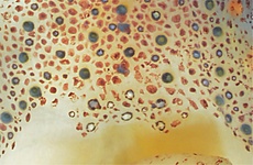
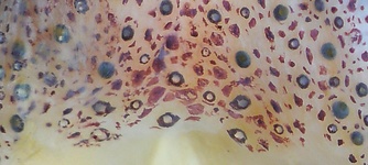
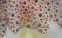
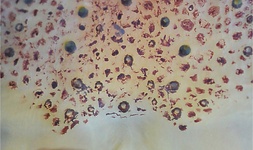
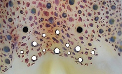
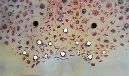
Tables. Ventral view of the funnel-groove region of Abraliopsis sp. NC4.
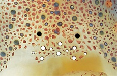
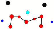
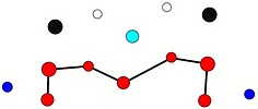
Top tier - Photograph of the preserved squid showing photophores of the funnel groove and surrounding tissue. Middle tier - In the photograph, white dots are placed over the "white" photophores of the funnel groove. Black dots are placed over the two most posteromedial "red" photophores of the ventral head to mark the anterolateral edges of the funnel groove. Bottom tier - Same pattern of dots as in photographs but dots are colored. Colored dots, lines - Aid in comparison of patterns between species. Open dots - Represent photophores that are either of uncertain status as "white" photophores or have variable presence. Images by R. Young. Comparison of white photophore patterns for all Abraliopsis species can be found here.
| Mature male, 20 mm ML | Mature female, 24 mm ML | Mature female, 26 mm ML. |
|---|---|---|
 |  |  |
 |  |  |
 |  |  |
| Mature female, 29 mm ML | Mature female, 32 mm ML |
|---|---|
 |  |
 |  |
 |  |
Comments: Summary of photophore distributions. (photophore definitions found here)
ARMS:
Medial Arm IV Series - Continuous; reaches about 2/3 of arm length.
Lateral Arm IV Series -Continuous; reaches arm tip.
Lateral Membrane Series of arms IV - Reaches about 2/3 of arm length.
Arm III Series - Continuous; nearly reaches the arm tip.
HEAD - Typical scattered pattern.
Median Head Series - Absent.
Median Head Sector - Scattered photophores. At 20 mm ML 3-5 red photophores, tendency for midline alignment. At 26 mm ML 3 red photophores, tendency for midline alignment.
Medial Head Series - Fairly arbitrary designation.
Lateral Head Sector - Scattered photophores. At 20 mm ML zero red photophores. At 26 mm ML one red photophore.
Lateral Head Series - Well defined.
Window Sector - Few scattered blues at either end. Otherwise without photophores.
1st Lateral Window Series - At 20 mm ML, two postertior segments with 2 red photophores each, one anterior segment of 5 red photophores at 20 and 26 mm ML.
2nd Lateral Window Series - Not present. At 20 mm ML, 0-1(questionable) red photophore; at 26 mm ML one red photophore.
Eyelid Series - Red photophores only on ventral half of eyelid. Posterior Eyelid Series with one or two large blue photophores displaced slightly posteriorly from eyelid series, if two, with the more ventral larger and more posterior. Formula: RbBB at 20 mm ML; RbBB at 24 (F); RbbBB at 26 mm ML; RbBbB at 29 mm ML; RbBB at 32 mm ML.
Occipital Series - Rbbb at 19 mm ML; RbbR at 20 mm ML; RbbR at 24 mm ML; RbBbR at 26 mm ML; RbRbR at 29 mm ML; RbbR at 32 mm ML.
Lateral Funnel-groove Series - Three red photophores at 20 mm ML, four at 26 mm ML.
Olfactory Photophore - Red at 26 mm ML, blue at 29 and 32 mm ML.
White Patch (funnel groove). Moderate Pattern (red, green, blue + yellow with size).
FUNNEL: Patches not well demarcated.
Medial Patch - Scattered red photophores.
Lateral Patch - Two red photophores at 20 and 26 mm ML..
MANTLE - Typical scattered pattern with:
Median Sector - Scattered blue photophores in anterior third of mantle tends to obscure central bare strip anteriorly.
Medial Mantle Series - Recognizable but irregular.
1st Lateral Mantle Sector - Recognizable, with distinctly more dense distribution of photophores than in 2nd Lateral Mantle Sector.
Lateral Mantle Series - Detectable and linear (red photophores).
Mantle-angle Series - Dectectable and linear but with usual anterolateral bend.
Comments
The linear tendency of photophores of the head and mantle are more apparent in this species than in Abraliopsis sp. B or A. pacificus.
The NC species were frozen prior to fixation which makes detection of photophore types, arm membranes and trabeculae difficult and, probably, with more errors in description.


