
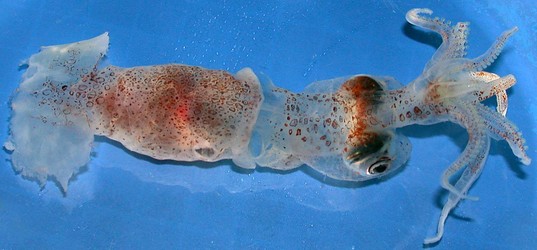
Figure. Dorsal view of P. levimana, neotype, 60 mm ML, immature female. Photograph by R. Young.
- Arms
- Arm suckers in two, closely packed series on arms I-III.
- Arms IV more fragile and elongate than other arms.
- Arm suckers with ca 10-15 broad, truncate to triangular teeth distally becoming low, rectangular teeth proximally. Holotype described as having truncate teeth all around the aperature.
- Arm I suckers smaller that lateral arm suckers.
- Arm IV smallest, 0.4 mm (outer rings), 0.23 aperatures.
- Large suckers of lateral arms with outer rings 0.8 mm, aperatures 0.5 mm in diameter.
- Arms with low protective membranes, often difficult to detect.
- Tentacles
- Stumps of tentacles thick, muscular (see above figure).
- Distal third of club with suckers in four series decreasing to one near tip.
- Suckers end prior to club tip which forms a short cone covered with chromatophores.
- Proximally suckers not as regularly arranged, appear mostly in three series, decreasing to a single sucker at base and preceeded proximally by a "carpus."
- "Carpus" consists of 5 suckers in single zig-zag series; suckers mostly larger than suckers of club proper and with relatively larger aperatures.
- Club suckers with broad outer rings and narrow aperatures; apparently with smooth inner rings.
- Entire club, including "carpus,"bounded by think protective membranes which apparently join distally after terminal sucker but prior to tip.
- Head
- Eyes large but laterally compressed; short, silvery rostra present on ventral tips of eyes.
- Eyes bulge greatly from head laterally and ventrally, but encapsuled in thick gelatinous layer along with lateral sides of head (see figures).
- Short brachial pillar estends from head to base of brachial crown.
- Broad, deep excavation between funnel adductor muscles.
- Bridles extend to mid-region of head, nearly at level of eye rostra.
- Olfactory organs on long slender stalks posterolateral to eyes.
- Mantle
- Mantle thin but muscular.
- Mantle terminates at midlevel on fins.
- Fins and tail
- Anterior lobes present, posterior lobes uncertain due to damage.
- Thick, triangular conus broken off at posterior edge of fins; tail missing.
- Photophores
- Photophores absent.
- Pigmentation
- Pigment in large reddish-brown chromatophores. Mantle more heavily pigmented than the head.
- Viscera
- Digestive gland oblique to body axis.
- Measurements
- Specimen no. 1 (immature female): ML - 60 mm, MW - 13 mm, FW - 36 mm, FL - 17 mm , HL (nuchal cartilage to posterior end of brachial pillar - 16.5 mm; Brachial pillar width - 9 mm, Branchial pillar depth - 7 mm, Head width (across eyes) - 14, Neck width - 13.5 mm, Neck length (collar to posterior eye margin) - 8 mm, Eye width -10 mm, Eye depth - 12.5 mm, Head depth (midline at posterior edge of eyes) - 12 mm, Lens diameter - 5 mm .
- Specimen no. 2 (sex ?): ML - 70 mm (estimate), Club length - 11 mm, Club width - 2.2 mm, Diameter outer ring of large suckers of club proper - 0.3 mm, aperture diameter - 0.06 - 0.08, Large carpal suckers - 0.04 mm, aperture - 0.14-0.23 mm.
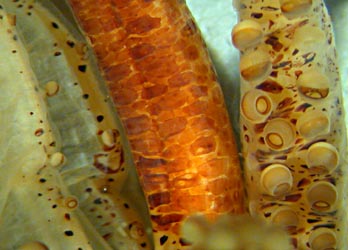
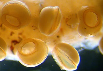
Figure. Arms and arm suckers of P. levimana. Left - Oral view of the bases of left arm IV, right arm IV, tentacle stalk, right arm III. Note the difference in sucker sizes and sucker packing between arms III and IV and the robust nature of the tentacle. Right - Oral and lateral views of large arm suckers. Note the easily recognized teeth. Photographs by R. Young.
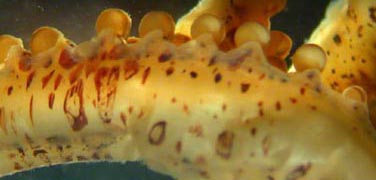
Figure. Lateral view of one of arms I-III of P. levimana, neotype, showing the low protective membrane near the arm base. Photograph by R. Young.
Scanning electron micrographs of the arm suckers can be seen here.
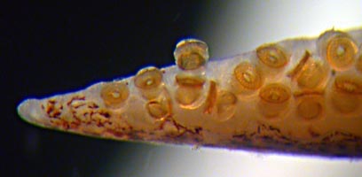
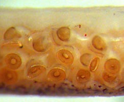
Figure. Oral views of portions of the tentacular club of P. levimana, 70 mm ML. Left - Club tip without any special modification of sucker arrangement (i.e. no termianal pad). Right - Proximal region of club. Note the variation in the sizes of the sucker aperatures. Photographs by R. Young.
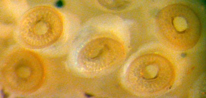
Figure. Oral view of club suckers, P. levimana, 70 mm ML. Pegs prominent in outer rings but teeth appear to be absent from inner ring. Photograph by R. Young.
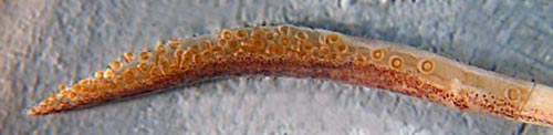
Figure. Oral view of the tentacular club of P. levimana, ca. 70 mm ML. Photograph by R. Young.
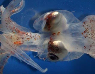

Figure. Head of P. levimana, neotype, 60 mm ML. Left - ventral view. Note the lateral compression of the eyes. Right - Dorsal view. Photographs by R. Young.
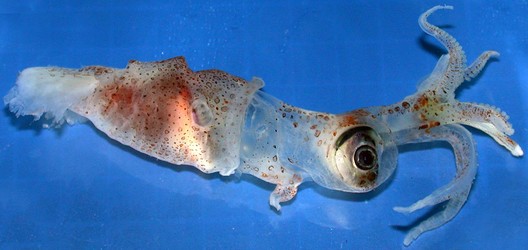
Figure. Side view of P. levimana, neotype, 60 mm ML. Note the oblique digestive gland and the laterally compressed and ventrally projecting eyes. Photograph by R. Young.
Comments
The above description is mostly taken from Young, et al. (2006).


