- Arms
- Arms in females (see drawing) lack enlarged suckers.
 Click on an image to view larger version & data in a new window
Figure. Oral view of arm I of O. agassizii, mature female. Drawing from Villanueva, et al. (2002).
Click on an image to view larger version & data in a new window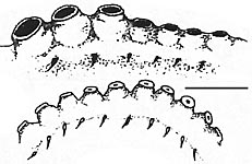
Figure. Side views of arm suckers of O. agassizii. Top - Proximal portion of an arm of a mature male showing enlarged suckers, neotype, 42 mm ML. Bottom - Proximal portion of an arm of a mature female, same specimen as previous drawing. Drawing from Villanueva, et al. (2002).
- Arms in females (see drawing) lack enlarged suckers.
- Funnel
- Funnel organ inverted V-shape.
- Funnel organ inverted V-shape.
- Gills
- 7 primary lamellae (rarely 6 or 8).
- 7 primary lamellae (rarely 6 or 8).
- Optic lobes and nerves
- Optic lobes kidney-shaped.
- 2-4 optic nerve bundles penetrate white body.
Click on an image to view larger version & data in a new window
Figure. Ventral view of the eye, optic nerve bundles, white body and optic lobe of a maturing female of O. agassizii, 33 mm ML. Drawing from Villanueva, et al. (2002).
- Digestive tract
- Digestive gland unilobular.
- Posterior region of esophagus expanded into crop.
- Intestine length about 1.5 times that of esophagus; intestine forms an S-shpape.
Click on an image to view larger version & data in a new window
Figure. Lateral view of the digestive system of O. agassizii, mature male, ca. 50 mm ML. Drawing from Villanueva, et al. (2002)
- Beaks
- Lower beak: rostrum pointed, hood small, wings long and narrow; lateral walls without folds or ridges.
Click on an image to view larger version & data in a new window
Figure. Lateral view of beaks of O. agassizii, mature male, 35 mm ML. Left - lower beak. Rght - Upper beak. Drawings from Villanueva, et al. (2002).
- Male reproductive system
- Accessory gland complex dominated by gland 3 (?).
- Penis short.
- Spermatophore gland complex convoluted.
- Spermatophores ellipsoidal, 0.7-1.6 mm in length.
Click on an image to view larger version & data in a new window
Figure. Reproductive tract of a mature male of O. agassizii. Drawing from Villanueva, et al. (2002).
- Female reproductive system
- Proximal oviduct long, distal oviduct short to moderate length.
- Proximal portion of oviducal gland colorless and slightly shorter than dark-colored distal portion.
- Drawings below are eggs from the ovary (left), the proximal oviduct (middle) and the distal oviduct (right).
Click on an image to view larger version & data in a new window
Figure. Reproductive tract of a mature female of O. agassizii. Drawing from Villanueva, et al. (2002).
Click on an image to view larger version & data in a new window
Figure. Eggs from the ovary and oviduct of O. agassizii. Drawings from Villanueva, et al. (2002).
- Shell
- U-shaped with short, flaring wings.
- Saddle with outer surface concave, inner surface convex.
- Wings flattened, termination varies from spike-like to broad, pointed end.
There are two major variations in shell morphology: A - Type A of Villaneueva et al. (2002) - Wing terminates in a spike that extends from a shoulder with a distinct shelf. B - Type B of Villaneueva et al. (2002) - Wing terminates in a broad, pointed end, shoulder without distinct shelf.
Click on an image to view larger version & data in a new window
Figure. Shell of O. agassizii. A- Ventral (top), dorsal (middle) and side (bottom) views of Type A shell. B- Ventral (top), dorsal (middle) and side (bottom) views of Type B shell. Drawings from Villanueva, et al. (2002).
Click on an image to view larger version & data in a new window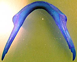
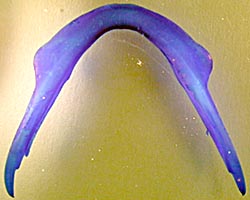
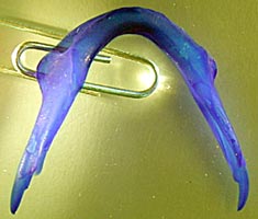
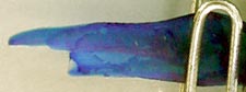
Figure. These photographs of stained shells of O. agassizii are of type A. Upper left - Ventral view. Upper right - Dorsal view. Lower left - Oblique view. Lower right - Lateral view of wing. Photographs by R. Young.
- Pigmentation
- Skin reddish-brown in preservation. Areolae often difficult to see depending on condition of animal; present on head and proximal 2/3 of all arms. Rows on dorsal arms extend onto head and mantle contain 12 spots; arms II and III each have 8-12 spots; arms IV each have 6 spots with proximal spot enlarged.
Click on an image to view larger version & data in a new window
Figure. Dorsal (left) and ventral (right) views of an immature female of O. agassizii, approximately 25 mm vental mantle length showing arrangement of areolae. Drawings from Villanueva, et al. (2002).
- Measurements
Opisthoteuthis agassizii: Description Continued
Richard E. Young, Michael Vecchione, and Roger VillanuevaReferences
Villanueva, R., Collins, M., Sanchez, P. and N. Voss. 2002. Systematics, distribution and biology of the cirrate octopods of the genus Opisthoteuthis (Mollusca, Cephalopoda) in the Atlantic Ocean, with description of two new species. Bulletin of Marine Science 71(2):933-985.
About This Page
Drawings printed with the Permission of the Bulletin of Marine Science.

University of Hawaii, Honolulu, HI, USA
National Museum of Natural History, Washington, D. C. , USA
Instituto de Ciencias del Mar (CSIC), Barcelona, Spain
Page copyright © 2003 , , and
 Page: Tree of Life
Opisthoteuthis agassizii: Description Continued
Authored by
Richard E. Young, Michael Vecchione, and Roger Villanueva.
The TEXT of this page is licensed under the
Creative Commons Attribution-NonCommercial License - Version 3.0. Note that images and other media
featured on this page are each governed by their own license, and they may or may not be available
for reuse. Click on an image or a media link to access the media data window, which provides the
relevant licensing information. For the general terms and conditions of ToL material reuse and
redistribution, please see the Tree of Life Copyright
Policies.
Page: Tree of Life
Opisthoteuthis agassizii: Description Continued
Authored by
Richard E. Young, Michael Vecchione, and Roger Villanueva.
The TEXT of this page is licensed under the
Creative Commons Attribution-NonCommercial License - Version 3.0. Note that images and other media
featured on this page are each governed by their own license, and they may or may not be available
for reuse. Click on an image or a media link to access the media data window, which provides the
relevant licensing information. For the general terms and conditions of ToL material reuse and
redistribution, please see the Tree of Life Copyright
Policies.


