
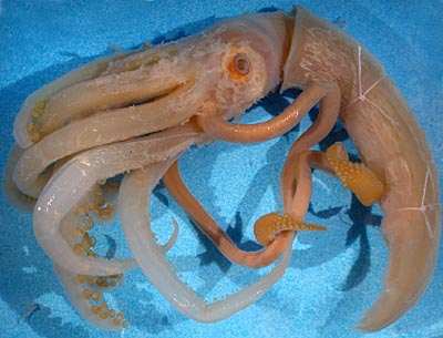
Figure. Lateral view of the specimen of N. thielei that most of the following description is based on, 100 mm ML, N. Pacific.
- Arms
- Arms I shortest; arms II-IV subequal.
- Largest suckers of arms I-III approximally equal in size (2.7 mm diam.); largest suckers of arms IV much smaller (1.1 mm diam.)(See photograph on below.).
- Delicate protective membranes without obvious trabeculae present on arms (badly damaged in specimen examined).
- Tentacles
- Locking apparatus
- "Carpal" locking suckers and knobs absent (see photograph at right showing absence of a locking apparatus at the base of the manus); locking suckers and knobs along tentacle stalk absent.
- Manal locking suckers present; evenly but broadly spaced along dorsal margin of proximal-manus in single series (see photograph on species page); difficult to recognize from normal manal suckers proximally and distally on present specimen. Locking knobs not seen.
- Manus
- Distal-manus with abruptly larger suckers than proximal-manus (about 2-4 suckers are of intermediate size).
- Club expanded, maximum width across proximal-manus ca 8% of ML.
- Suckers of proximal-manus very small, smaller at proximal end (0.2 mm proximally, 0.25mm distally); about 35 suckers across maximum width of proximal-manus near distal end; about 25 suckers across proximal-manus near proximal end where club first expands.Suckers have round aperatures. The above photographs show three different regions of the proximal-manus (left - distal end; middle - slightly more proximal; right - well proximal).
- Dactylus and terminal pad
- About 5 suckers initially across distal-manus but shortly becomes four regular series as it grades into dactylus and throughout dactylus.
- Terminal pad (=distal locking-apparatus) with 16-20 suckers but difficult to determine where dactylus suckers end and pad suckers begin. Pad suckers form a circle with a central space and the sides of the circle are composed of two series of suckers.
- Head
- Eyes large, diameter 18% of ML.
- Buccal connectives attach to dorsal margins of arms IV.
- Occipital folds absent.
- Olfactory organ on short stalk.
- Beaks not examined.
- Mantle
- Mantle wall thick but spongy; arms extremely delicate.
- Fins
- Fins separate throughout length; separation increases anteriorly; anteriorly attach to sides of mantle. (In the title photograph, the long fins reach to the anterior most string tied around the mantle.)
- Fins slender, combined width 30% of ML.
- Posterior lobes probably present (damaged); without anterior lobes.
- Photophores
- Photophores absent.
- Gladius
- The morphometrics of the gladius have been reported by Toll, 1982: Gladius length - 105.6 mm; free rhachis length - 12.2 mm; free rhachis width - 4.3 mm; maximum gladius width (across vanes) - 8.0 mm; conus length - 2.0 mm.
- Pigmentation
- Buccal membrane unpigmented; no pigment remains on animal surface. Remnants of thick white tissue present on aboral surfaces of arms, surfaces of head and thin traces on mantle (see title photograph).
- Measurements
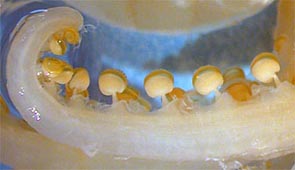
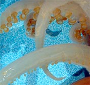
Figure. Left - Lateral view of mid-region of arm II of N. thielei, N. Pacific. Right - Ventral-oblique view of the two arms IV and several more dorsal arms; same specimen. Photograph by R. Young.
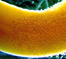
Figure. Oral view of the proximal end of the proximal-manus of N. thielei, 100 mm ML, N. Pacific.
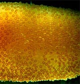
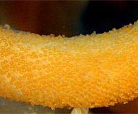
Figure. Oral view of portions of the club of N. thielei, 100 mm ML, N. Pacific. Left - Region where the proximal- and distal-manus regions meet. Middle - Distal region of the proximal-manus. Right - Proximal region of the proximal-manus. Photograph by R. Young.
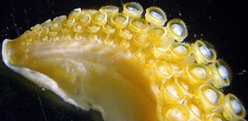
Figure. Oral view of the dactylus of N. thielei, N. Pacific. Photograph by R. Young.

Figure. Ventral-oblique view of the head of N. thielei, N. Pacific. Photograph by R. Young.
| Specimen | USNMNH 730705 |
| Sex | Female |
| Mantle length, mm | 100 |
| Mantle width | 19 |
| Head length | 26 |
| Head width | 23 |
| Arm length I, Left / Right | 58 / - |
| Arm length II | 76 / 57 |
| Arm length III | 84 / - |
| Arm length IV | 87 / 93 |
| Club length | 63 / 59 |
Comments
The gladius was previously removed by R. Toll, but it remains in the jar with the squid. This squid is a female.

