


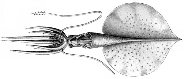
Magnapinna talismani
Michael Vecchione and Richard E. Young
Introduction
Magnapinna talismani was originally described from a single damaged specimen that came from south of the Azores, North Atlantic very near the capture site of Magnapinna sp. B and the paratype of M. atlantica. On this page we will summarize the original text, include illustrations that accompanied the text along with photographs and comments from our examination of the holotype. Then we present the text of the original description of M. talismani, first described as Chiroteutopsis talismani. The identy of M. talismani is uncertain. It is similar to M. atlantica and Magnapinna sp. C and may ultimately prove to be conspecific with these species.
Brief diagnosis:
A Magnapinna with ...- slender proximal-tentacles that lack suckers and glandular structures.
Characteristics
- Arms
- Suckers on proximal-arm in two series and closely packed.
- Proximal-arm suckers with smooth inner rings.
- Arms badly damaged.
Click on an image to view larger version & data in a new window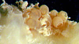
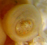
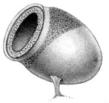
Figure. Proximal-arm suckers of M. talismani, holotype. Left - Side view of a portion of the proximal-arm. Middle - Oral view of an arm sucker. Photographs by R. Young. Right - Oblique view of an arm sucker. Drawing from Fischer and Joubin (1907).
- Tentacle
- Tentacles badly damaged; long, thin and cylindrical; much thinner than the arms at their base.
- In contrast to the original description and illustration (see drawing above), no evidence of a discreet club is present.
- No disinct transition between proximal- and distal-tentacle.
- Distal-tentacle apparently with numberous, small suckers (photograph and drawing below).
Click on an image to view larger version & data in a new window
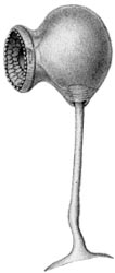
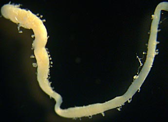
Figure. Tentacles of M. talismani, holotype. Left - Ventral (?) view of the brachial crown showing slender proximal-tentacle adjacent to much thicker arms. Photograph from Vecchione and Young (submitted), modified. Middle - Side view of a tentacle sucker. Drawing from Fischer and Joubin (1907). Right - Appendage free in jar with holotype, apparently a portion of the tentacle. Note what appear to be small suckers on the badly abraided appendage. Photographs by R. Young
- Funnel
- Funnel locking-apparatus with oval depression and possible tragus (from F&J). Both funnel locking-cartilages are distorted in the holotype and the presence or absence of a weak tragus is uncertain but unlikely.
Click on an image to view larger version & data in a new window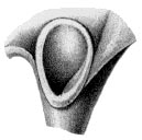
Figure. Frontal view of the funnel locking-apparatus of M. talismani, holotype. Drawing from Fischer and Joubin (1907).
- Mantle
- Mantle length 61 mm (from F&J).
- Mantle length 61 mm (from F&J).
- Fins
- Ventral surface of fins covered with white nodules (from F&J).
- Fin length - 54 mm; fin width - 53 mm (from F&J).
- No tail present.
- Apex angle of fins nearly 90°. Click on an image to view larger version & data in a new window
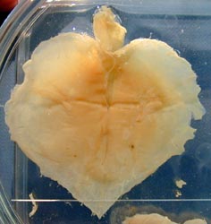
Figure. Dorsal view of the fins and mantle of M. talismani, holotype. A glass slide has been placed over the posterior tip of the fins to keep them flat. Photograph by R. Young.
- Gladius
- Gladius not removed. The length of the gladius cannot be determined as the anterior end is frayed indicating that some of it may be missing.
Comments
Very little new information can be derived from our examination of the holotype. Information from the original description that we did not confirm is marked "from F&J" above. This squid was badly damaged when captured and has not survived well in preservation. The poor condition of the holotype which has lost most species-level characters makes comparisons with other species difficult.
Nomenclature
This M. talismani was originally placed in the genus Chiroteuthopsis which is a junior synonym of Mastigoteuthis. Mastigoteuthis talismani was placed in Magnapinna by Vecchione and Young (2006).
Distribution
Type locality: 34°46'N, 36°11'W (south of the Azores, North Atlantic). The holotype was caught in an open bottom trawl, that fished to a depth of 3175 m.
Text of the Original Description
TT 78-55121
From: Exped. Scient. du Travailleur & Talisman. Vol. 8, 1907 pp. 342-345.
CEPHALOPODS
By: Fischer, H. and L. Joubin
Translated from French by: Margaret Saidi
Edited by: Azzedine Azzouz
Translated for The National Science Foundation, Washington, D.C. and The Smithsonian Institution by The Agence Tunisienne de Public-Relations Tunis-Tunissie.
-1979-
SUBFAMILY CHIROTEUTHIDAE GRAY, 1849 (l9)
GENUS CHIROTEUTHOPSIS PFEFFER. 1900 (47).
17. Chiroteuthopsis Talismani, nov. sp.
(Pl. XXV, figs. 1 to 4)
Talisman. - Dr. 118. August 10, 1883. Depth 3175 m.
Lat. N. 34°46'. Long. W, 36° 11'. - South of the Azores. -
Bottom of pumice stone.
Observations. A single specimen of this singular species has been caught; it is probable that it does not come from the bottom, but that it was caught between two waters when the trawl-net was raised, as it is a distinctly bathypelagic cephalopod. It was unfortunately greatly damaged by its stay in the net, and we can therefore give only a very incomplete description of it. The tentacles have, among other things, been twisted and broken, and it is impossible to measure their length. The ends of the arms have undergone the same fate; only their bases are more or less intact.
What characterizes this animal right from the first examination is the enormity of its fin, approximately round, slightly pointed at the posterior extremity of the body. In proportion to the general size of the body, this fin is larger than in any other cephalopod of the family of the Chiroteuthidae. The visceral sac, on the other hand, is very small. It has the form of a very pointed cone, three-fourths of the length of which lie against the fin. This cone is surmounted by the upper part of the body, which projects, in the form of a semitransparent cylinder, directly into the head. The latter is not distinct from the body; it has two eyes at its summit. They do not appear to have been large, but their bad condition makes it impossible to be clear.
The crystalline lenses of these eyes formed the two upper angles of the head, and it is between them, through a very narrow very low neck, that the tentacular and brachial crown is attached.
This crown seems to have had the general form of that of all the Chiroteuthidae; what remains is not sufficient to enable us to give a table of measurement of the arms. Figure 1 of plate XXV is a reconstruction of the animal, the length of the arms being indicated approximately, according to the debris which remained attached to the stumps. The skin of the arm bases, on their buccal face, and the membranes surrounding the lip, are dark purple in color. It is still possible to see, on two rows, some suckers, which appear to have been yellow. The circular lip, with a wide opening, surrounds relatively large mandibles; this lip is yellow, tending toward the violet tint of the membranes which surround it.
The tentacles formed a twisted mass from which it was possible to isolate only fragments. We were nonetheless able to find the tentacular plate of one of these, still bearing a few excessively small suckers, posed on a lang, thin peduncle; these suckers appear to have been very numerous.
We give below the measurements it was possible to take from this specimen; they are very incomplete because of the almost total destruction of the appendages. It is probable that the long period spent in alcohol has reduced the animal's size; there exists, in fact, a sketch made immediately after its capture by C. Brongniarf. This sketch, which is larger than the sample represented, gives, after deduction of the indicated enlargement, a decrease of approximately one-quarter.
Distance from the tip of the fin to the mouth -- 90 mm
Height of the fin -- 54
Width of the fin -- 53
Distance from the palleal edge to the tip of the fin -- 61
Distance from the neck to the upper edge of the fin -- 26
Diameter of the head at the level of the eyes -- 9
According to Brongniarf’s sketch, the general color of the body and of the fin is pale fleshy rose; that of the inner surface of the arm is dark purple, while that of the outline of the mouth is yellowish brown.
We shall now review certain details of this cephalopod. The fin is approximately round, soft, scattered with large isolated chromatophores; a few whitish nodules on the ventral face may be the remains of light organs; it is impossible to be certain because of the state of the specimen. These chromatophores are abundant on the dorsal face, rarer on the central face, The fin ends on the bottom in a short point determined by the extremity of the plume. On the top, behind the insertion of the body, there is a deep notch in the fin. The musculature of this organ is powerful, and this cephalopod must be a strong swimmer.
The body can be broken down into two parts. One is an upper, cylindrical part extending from the brachiai crown to the palleal edge; the other is conical, extending from the palleal edge to the tip of the fin.
The upper part is smooth dorsally; ventrally the eyes lie in the upper part, the funnel in the middle, and at the bottom the visceral mass, including the rectum, the ink sac, and the tip of the female genital glands, particularly two white, oval, slanted nidamentary glands.
The gullet is well developed. It bears, on both sides of its lower opening, deep, projecting, very highly developed adhesive pits. Their lower edge is formed of a rounded cartilage with a well-delineated contour. The contour or the upper edge is less distinct, blending gradually into the skin of the siphon. A strong ligament attaches the bottom of the pit to the visceral mass. Outside and above this ligament, the lateral valve of the gullet is well developed. It was impossible to see exactly the inner structure of the oval pit. It appears, however, to contain a tragus. Similarly, it is impossible to say whether there is a valvule in the gullet.
The palleal edge is soft; its contour appears to be circular, without sinuosity, on the ventral edge. On the back, it rises slightly higher, but bears no nuchal cartilage.
The conical part of the body is short; it exceeds the edge of the fin by about nine millimeters. Beyond, it thins rapidly and lies so completely against the fin that it appears to be a part of it. The upper part of this conical region is attached to the notched region of the fin by two well-developed triangular membranes.
The eyes are in bad condition; they seem to be little-developed, It is impossible to say whether they possessed light organs.
The arms are in a regular crown; only the bases are more or less intact. They appear to have been of approximately the same size. It is impossible to measure their length, since most of them are now no more than debris, They bear two rows of suckers which can be seen in place at the bases of two or three arms. They are ovoid, pierced with an orifice surrounded by a well-developed cornate circle with platelets disposed over three or four rows, each surmounted by a small tubercle. The peduncle is very short; the suckers appear to have been very close to one another.
The tentacles are thin and cylindrical. They seem to have been eight to ten centimeters long, in so far as can be judged by assembling their fragments. They end in a narrow paddle, eight to ten millimeters long, bearing a large number of very small suckers, with a very long peduncle. They are approximately spherical, with two projecting heels around the insertion of the peduncle, which is embedded between them. The circular orifice is provided with three rows of plates, the smallest at the edge, provided, with a tubercle, and the larger within without tubercle. The bright yellow chitinous coating covers the entire interior of the cavity of the sucker.
This cephalopod belongs distinctly to the Chiroteuthidae; it belongs more particularly to the genus Chiroteuthopsis, created specially by Pfeffer for a species, Ch. Grimaldii Joubin (30), initially placed by the author in the genus Chiroteuthis (47), The genus of Pfeffer now therefore Includes two species.
References
Fischer, H. and L. Joubin (1907). Expéditions scientifiques du TRAVAILLEUR et du TALISMAN. Céphalopodes, 8: 313-353.
Vecchione, M. and R. E. Young. 2006. The squid family Magnapinnidae (Mollusca; Cephalopoda) in the North Atlantic with a description of Magnapinna atlantica, n. sp. Proc. Biol. Soc. Wash. 119 (3): 365-372.
Title Illustrations

| Scientific Name | Magnapinna talismani |
|---|---|
| Location | North Atlantic Ocean |
| Reference | Fischer, H. and L. Joubin (1907). Expéditions scientifiques du TRAVAILLEUR et du TALISMAN. Céphalopodes, 8: 313-353. |
| Specimen Condition | Dead Specimen |
| View | Ventral/dorsal |
| Size | 61 mm ML |
| Type | Holotype |
About This Page

National Museum of Natural History, Washington, D. C. , USA
University of Hawaii, Honolulu, HI, USA
Page copyright © 2016 and
 Page: Tree of Life
Magnapinna talismani .
Authored by
Michael Vecchione and Richard E. Young.
The TEXT of this page is licensed under the
Creative Commons Attribution-NonCommercial License - Version 3.0. Note that images and other media
featured on this page are each governed by their own license, and they may or may not be available
for reuse. Click on an image or a media link to access the media data window, which provides the
relevant licensing information. For the general terms and conditions of ToL material reuse and
redistribution, please see the Tree of Life Copyright
Policies.
Page: Tree of Life
Magnapinna talismani .
Authored by
Michael Vecchione and Richard E. Young.
The TEXT of this page is licensed under the
Creative Commons Attribution-NonCommercial License - Version 3.0. Note that images and other media
featured on this page are each governed by their own license, and they may or may not be available
for reuse. Click on an image or a media link to access the media data window, which provides the
relevant licensing information. For the general terms and conditions of ToL material reuse and
redistribution, please see the Tree of Life Copyright
Policies.
- First online 16 August 2005
- Content changed 08 February 2007
Citing this page:
Vecchione, Michael and Richard E. Young. 2007. Magnapinna talismani . Version 08 February 2007 (under construction). http://tolweb.org/Magnapinna_talismani/52212/2007.02.08 in The Tree of Life Web Project, http://tolweb.org/


