


Abraliopsis sp. NC1
Richard E. Young
Introduction
Abraliopsis sp. NC1 is presently known only from waters near New Caledonia. Females reache a length of at least 28 mm ML.
Brief diagnosis:
An Abraliopsis (Micrabralia) ...
- with distal third/half of arms IV bare of hooks and suckers (both sexes).
- without keels and carpal flaps on tentacular clubs.
- with largest club hook of ventral series <1.5 X height of dorsal counterpart.
- without base of arms I enlarged in males.
Characteristics
- Arms
- Hectocotylus (right arm IV): Ventral flap long, wide; dorsal flap shorter, wide, offset distally. Hooks extend to proximal end of ventral flap.
- Arms I-III: Females with well-developed trabeculate protective membranes on ventral margins of arms I-III; trabeculae slender; tubercules absent. Dorsal margins virtually without membranes or trabeculae. Arms IV with low, thick membranes on armature-bearing proximal arms. Arms I without enlarged hooks.
- Arms IV: Males and females lack armature on distal 1/3 to 1/2 of arms.
Click on an image to view larger version & data in a new window
Figure. Side view of Arms IV of Abraliopsis sp. NC1, female, 28 mm ML, showing bare distal half of arms. Photographs by R. Young.
- Arms I-III males:
- Arms I - with large trabeculae on both margins, those of ventral margin longer and broad. Trabeculae bear tubercules. Tubercules also on oral base of arm and, probably, on some areas of oral arm surface between hooks. (Squid examined had been frozen prior to fixation which makes tubercules difficult to detect.)
- Arms II-III - Large trabeculae on ventral margins only. Tubercules not apparent on trabeculae or arm bases. Base of arm II not swollen.
Click on an image to view larger version & data in a new window
Figure. Oral view of arms IV of Abraliopsis sp. NC1, mature male, 20 mm ML. Photograph by R. Young.
Click on an image to view larger version & data in a new window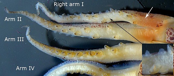
Figure. Oral views of arms I-III and dorsal view of portion of arm IV of Abraliopsis sp. NCI, male, 27 mm ML. White arrow points to tubercules on oral base of arm I. Insert (lower right) is an enlargement of a portion of arm I (black arrow) showing tubercules.
- Hectocotylus (right arm IV): Ventral flap long, wide; dorsal flap shorter, wide, offset distally. Hooks extend to proximal end of ventral flap.
- Tentacles
- Club without keel or carpal flap.
- Club hooks not greatly different in size between ventral and dorsal series (large ventral hooks < 1.5 times length of dorsal counterparts).
- Claw of large club hooks without strong lateral compression in cross-section.
- Head
- Occipital folds: Although poorly formed due to freezing prior to fixation, cresent membrane between folds 3 and 4 apparently well formed.
- Occipital folds: Although poorly formed due to freezing prior to fixation, cresent membrane between folds 3 and 4 apparently well formed.
- Photophores
- Ocular photophores: 5 photophores, end members about 2.5-3 times diameter of middle photophore.
- Integumental photophores: Ventral head with red photophores in 3 series and ventral mantle in 6 series with scattered blue photophores between.
Click on an image to view larger version & data in a new window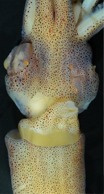

Figure. Ventral view of Abraliopsis sp. NC1, female, 26 mm ML. Left - Photograph of the preserved squid. The patterns can be difficult to detect in a quick look. Right - Drawing from the same photograph showing the distribution and approximate size of two classes of photophores (red dots - complex photophores, blue dots - non-complex photophores).
More information on the integumental photophores can be found here.
- Ocular photophores: 5 photophores, end members about 2.5-3 times diameter of middle photophore.
- Viscera
- Females with three receptacles for spermatangia, two dorsal-collar pockets adjacent to nuchal cartilage and one stellate pocket beneath the gladius. Pigment of receptacles visible externally. All receptacles generally contain spermatangia in mated females.
Click on an image to view larger version & data in a new window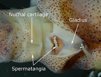
Figure. Dorsal view of the locations of the spermatangia receptacles in Abraliopsis sp. NC1, female, 26 mm ML. Top - In the photograph the squid has been bent ventrally to expose the nuchal cartilage and the associated dorsal-collar pockets. Left arrow points to the internal pigment of one dorsal-collar pocket. Right arrow points to pigment of the stellate pocket. Bottom - Mantle folded back to show spermatangia (white structures. On the left they are seen through the collar muscle), and the pigmented stellate pocket.
- Females with three receptacles for spermatangia, two dorsal-collar pockets adjacent to nuchal cartilage and one stellate pocket beneath the gladius. Pigment of receptacles visible externally. All receptacles generally contain spermatangia in mated females.
- Measurements and counts
NEC-1013, M018-68 (313)
NEC-1010, M011-40
NEC-1010, M011-40
NEC-1010, M011-40
NEC-1010, M011-40
NEC-1010, M011-40
NEC-1010, M011-40
Sex
Female, mated
Female, mated
Female, mated Female, mated
Male, mature
Male, mature
Male, mature
Mantle length
28
26
23
22
27
20
22
Head width index
-
-
-
-
-
Fin Length index
63.6
65.8
63.5
64.5
68.5
65
60.5
Fin width index
90.7
102.7
100
102.7
89.3
95
104.1
Arm Length index (R/L):
I
56.1/58.9
57.7/63.5
57.4/60.9
60.5/60.5
60.7/56.3
69/75
-/69.1
II
63.6/68.2
73.5/65
63.0/65.2
75/75.5
75.2/73.3
82.5/79.5
70.5/72.3
III
63.2/61.8
72.3/65
65.2/65.2
72.3/66.4
65.9/66.3
82.5/66.5
74.1/72.3
IV
76.1/77.1
74.6/76.5
82.6/82.6
102.2/95.6 77.8/85.6
89/91
82.7/81.8
No. arm hooks (R/L):
I
18/15 23/18
19/18
20/19
25/24
22/21
-/23
II
19/18
21/20
14/20
20/22
21/20
21/19
21/23
III
18/18
20/20
-/18
22/21
22/19
20/20
21/24
IV
10/10
8/9
9/9
10/10
12/13
13/14
11/11
Club length index (R/L)
22.9/22/9
26.9/-
24.8/26.1
27.3/32.7
-
-/29
25.9/25.9
Club hooks (D/V)(R:L)
4/4; 4/4
4/4; -
4/5; 3/3
-/4; 4/4
-
-; 3/4
3/3; 4/4
Carpal suckers (R/L)
4/4
4/-
-/-
4/3
-
-
-
sp.nc1f28club.300a.jpg)
Comments
In Abraliopsis sp. NC1, the tentacular club, the general photophore pattern and the unusual male modifications of arms I are similar to those of A. lineata. However the absence of swollen bases of arms II in males and the presence of bare distal arms IV separates it from A. lineata.
At present Abraliopsis sp. NC1 is distinctive in having 3 red photophores in the caret of the Median Head Series along with the 3 reds of the Medial Patch of the funnel and a single red in the Lateral Patch. Unfortunately, the details of the photophore patterns of A. lineata are unknown.
See the Abraliopsis (Micrabralia) page for comparisons among all species of the subgenus.
Distribution
Geographical distribution. At present Abraliopsis sp. NC1 is known from only two general localities. Most captures have been from northwest of New Caledonia near 18°S, 160°E, and one capture from a few degrees north of Samoa at 10°46'S, 172°34'W.
Figure. Distribution of Abraliopsis sp. NC1. White dots represent the capture localities. Base map from Google Earth.app.
Title Illustrations
| Scientific Name | Abraliopsis sp. NC1 |
|---|---|
| Location | Vicinity of New Caledonia |
| Specimen Condition | preserved |
| Sex | Female |
| View | Ventral |
| Size | 26 mm ML |
| Image Use |
 This media file is licensed under the Creative Commons Attribution License - Version 3.0. This media file is licensed under the Creative Commons Attribution License - Version 3.0.
|
| Copyright |
©

|
About This Page
University of Hawaii, Honolulu, HI, USA
Correspondence regarding this page should be directed to Richard E. Young at
Page copyright © 2016
 Page: Tree of Life
Abraliopsis sp. NC1.
Authored by
Richard E. Young.
The TEXT of this page is licensed under the
Creative Commons Attribution-NonCommercial License - Version 3.0. Note that images and other media
featured on this page are each governed by their own license, and they may or may not be available
for reuse. Click on an image or a media link to access the media data window, which provides the
relevant licensing information. For the general terms and conditions of ToL material reuse and
redistribution, please see the Tree of Life Copyright
Policies.
Page: Tree of Life
Abraliopsis sp. NC1.
Authored by
Richard E. Young.
The TEXT of this page is licensed under the
Creative Commons Attribution-NonCommercial License - Version 3.0. Note that images and other media
featured on this page are each governed by their own license, and they may or may not be available
for reuse. Click on an image or a media link to access the media data window, which provides the
relevant licensing information. For the general terms and conditions of ToL material reuse and
redistribution, please see the Tree of Life Copyright
Policies.
- First online 03 November 2013
- Content changed 03 November 2013
Citing this page:
Young, Richard E. 2013. Abraliopsis sp. NC1. Version 03 November 2013 (under construction). http://tolweb.org/Abraliopsis_sp._NC1/149528/2013.11.03 in The Tree of Life Web Project, http://tolweb.org/


